Mesothelioma Drug Development & Imaging: The Complete Guide to mRECIST, iRECIST & Imaging CRO Support
Mesothelioma Drug Development & Imaging:
The Complete Guide to mRECIST, iRECIST & Imaging CRO Support
A comprehensive clinical and operational guide for sponsors developing drugs in malignant pleural mesothelioma (MPM)
Understanding Malignant Pleural Mesothelioma: A Disease That Demands Precision
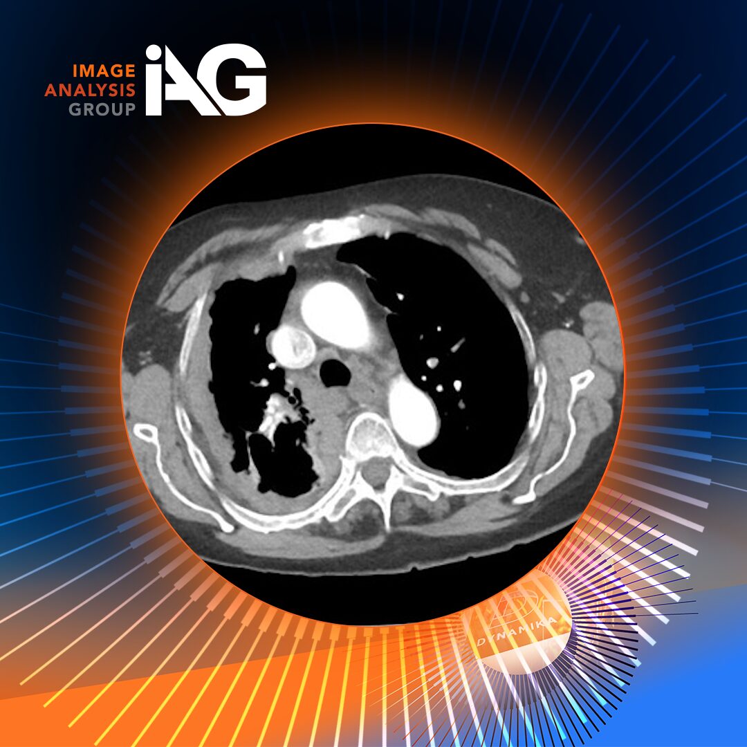
Malignant pleural mesothelioma (MPM) is one of oncology’s most challenging cancers – not only clinically, but from an imaging and trial design perspective. Arising from the mesothelial lining of the pleural cavity, MPM spreads in a diffuse, rind-like pattern along the pleural surfaces rather than forming discrete, spherical tumour masses. That single anatomical fact cascades into a series of complex challenges for everyone involved in a clinical trial: the radiologist reading the scan, the sponsor designing the endpoint, the CRO managing the imaging workflow, and ultimately the regulator evaluating the dossier.
The median survival for unresectable MPM remains under 20 months even with the most effective immunotherapy regimens currently approved. With relatively few active drug candidates in the pipeline and an orphan disease designation limiting large Phase III trial sizes, every imaging data-point carries outsized weight. A single protocol deviation in CT measurement methodology can jeopardise a primary endpoint. An inconsistent reader assessment can introduce bias that costs a drug its approval.
This article explains, in clinical and operational detail, exactly how imaging works in mesothelioma trials – the CT requirements, the measurement methodology of mRECIST, the parallel iRECIST track for immunotherapy studies, the eCRF module architecture that captures every data-point, and critically the role a specialist imaging CRO plays in ensuring that the imaging programme is not a weak link in an otherwise strong trial.
The MPM Drug Pipeline: What’s in Development and Why Imaging Matters
Mesothelioma drug development accelerated sharply between 2020 and 2026, driven by the success of immune checkpoint inhibitors (ICIs) and a renewed interest in novel targeted approaches. Understanding the pipeline helps frame why imaging endpoint methodology is so important: most active trials rely on radiographic response assessment as either a primary or key secondary endpoint.]
| Drug / Regimen | Mechanism | Phase | Primary Endpoint | Status |
| Nivolumab + Ipilimumab | PD-1 + CTLA-4 blockade | FDA Approved | OS (mRECIST) | First-line, 2020 |
| Pembrolizumab + Pemetrexed/Platinum | PD-1 + chemo (KEYNOTE-483) | FDA Approved | PFS, OS | First-line, 2024 |
| Durvalumab | PD-L1 blockade | Phase 3 | PFS (iRECIST) | Active enrolment |
| Volrustomig | PD-1 + CTLA-4 bispecific | Phase 3 | OS, PFS | Active |
| IK-930 (TEAD inhibitor) | Hippo/YAP pathway | Phase 1/2 | ORR (mRECIST) | NF2-mutant MPM |
| CAR-T (mesothelin) | Mesothelin-directed CAR-T | Phase 1 | Safety, ORR | Exploratory |
Source: OAE CDR 2025 (https://www.oaepublish.com/articles/cdr.2025.215) · Picone et al., Cancer Control 2025 (https://pmc.ncbi.nlm.nih.gov/articles/PMC11912172/)
The landmark CheckMate 743 trial demonstrated a median overall survival of 18.1 months with nivolumab plus ipilimumab versus 14.1 months with chemotherapy (HR 0.73). KEYNOTE-483 followed with pembrolizumab plus chemotherapy achieving an ORR of 52% versus 29%, with OS of 17.3 versus 16.1 months. Both trials share dependence on precisely conducted CT imaging assessments using mRECIST methodology. Measurement variability between readers has been shown to flip response category (PR vs. SD, or SD vs. PD) in a significant minority of cases, underscoring why BICR and a rigorous imaging CRO infrastructure are not optional additions – they are regulatory requirements for pivotal trials.
A single protocol deviation in CT measurement methodology can jeopardise a primary endpoint. In MPM, where the tumour grows like a rind rather than a sphere, imaging discipline is the difference between a successful trial and one that fails at the NDA stage.
CT Imaging in MPM: Techniques, Requirements and Challenges
Why Standard RECIST 1.1 Fails in Mesothelioma
RECIST 1.1 was designed for spherical or roughly spherical solid tumours — it measures the longest diameter of a lesion in the axial plane. MPM does not cooperate. The tumour wraps around the lung, grows bidirectionally along pleural surfaces, and — when responding — does not shrink centrally but instead thins its rind. Measuring the longest diameter of this geometry is uninformative. A pleural rind that has thinned from 25 mm to 15 mm in thickness represents a genuine and clinically significant response, yet might register minimal change on a standard longest-diameter measurement if the overall extent of disease is unchanged.
Modified RECIST 1.1 (mRECIST) was developed specifically to address this. Instead of measuring length, it measures perpendicular thickness of the pleural rind — the distance from the inner pleural surface to the lung or mediastinum, measured at right angles to the chest wall. This single methodological change makes the criterion genuinely sensitive to MPM biology, but it also introduces significant reader training requirements and protocol precision demands.
CT Protocol Requirements
The optimal CT protocol for MPM assessment requires multi-detector CT with intravenous contrast, with specific technical parameters that must be met at every timepoint:
| MANDATORY CT PROTOCOL PARAMETERS (mRECIST) |
| • Scanner type: Multi-detector CT (MDCT), 64-slice or greater |
| • Axial slice thickness: ≤2 mm (thicker slices underestimate pleural thickness) |
| • Sagittal/coronal slice thickness: ≤3 mm reconstructions |
| • Contrast: IV iodinated contrast, portal-venous phase (60–70 s delay) |
| • Coverage: Full thorax; must extend to L3 to capture subdiaphragmatic disease |
| • Reconstruction planes: Axial, sagittal, and coronal — all required |
| • Breath-hold: Single breath-hold preferred to minimise motion artefact |
| • Windows: Soft tissue, lung, and bone windows all available to reader |
Source: Picone C et al., Cancer Control 2025 – PMC11912172 (https://pmc.ncbi.nlm.nih.gov/articles/PMC11912172/)
Measurement Landmarks and the Perpendicular Approach
The radiologist reader must identify up to six target lesion sites across a maximum of three axial CT levels. Key constraints:
- Slices selected for measurement must be at least 1 cm apart in the z-axis
- Preferred levels: above the left atrium and below the aortic arch
- Each measurement must be perpendicular to the chest wall or mediastinum — not the longest diameter
- Minimum measurable size: 7 mm in the perpendicular direction
- Exception: if too thin to obtain 7 mm, a default value of 2 mm is applied (the TL-4 flag in the eCRF)
mRECIST – The Measurement Framework in Detail
Sum of Perpendicular Measurements (SPM)
The primary numeric output of each reading timepoint is the Sum of Perpendicular Measurements (SPM): the sum of perpendicular thickness at all target lesion measurement sites. The SPM at baseline establishes the reference. All subsequent SPMs are compared to the baseline SPM (for initial response) or the nadir SPM (for progression from best response).
SPM Worked Example – Baseline Timepoint
| Site | Location | Baseline (mm) | Week 12 (mm) | % Change | Flag |
| TL-1 | L pleural, above L atrium | 18.4 | 13.1 | −28.8% | — |
| TL-2 | L pleural, below aortic arch | 12.1 | 8.4 | −30.6% | — |
| TL-3 | R pleural, above L atrium | 22.7 | 15.9 | −29.9% | — |
| TL-4 | R pleural, below aortic arch | 2.0* | 2.0* | 0.0% | ⚑ 2mm default |
| TL-5 | R pleural, carina level | 15.9 | 11.1 | −30.2% | — |
| TL-6 | Mediastinal | 8.3 | 5.8 | −30.1% | — |
| SPM TOTAL | 79.4 mm | 56.3 mm | −29.1% | ⚠ Borderline SD/PR |
* TL-4 below 7 mm minimum — 2 mm default applied per mRECIST rules.
Source: Picone et al., Cancer Control 2025 (https://pmc.ncbi.nlm.nih.gov/articles/PMC11912172/)
Response Thresholds
| Category | Code | SPM Criterion | Additional Criteria |
| Complete Response | CR | No residual disease | Non-target resolved; no new lesions |
| Partial Response | PR | ≥30% decrease from baseline SPM | No new lesions; non-target not progressing |
| Stable Disease | SD | Neither PR nor PD criteria met | Reference: nadir SPM |
| Progressive Disease | PD | ≥20% increase from nadir + ≥5 mm absolute | New lesion = automatic PD regardless of SPM |
| Not Evaluable | NE | Insufficient data for assessment | Scan quality failure, missing timepoint, protocol deviation |
Source: Picone et al., Cancer Control 2025 · Revised mRECIST, J Thorac Oncol 2018
| ⚠ Critical Note: Borderline SD/PR Assessments The SPM example above (−29.1%) illustrates a borderline SD/PR assessment. These cases require adjudication by a third independent reader. Robust adjudication workflows — such as those built into DYNAMIKA™ — are critical for data integrity and regulatory acceptance. |
iRECIST – The Immunotherapy-Specific Track
The approval of immune checkpoint inhibitors for MPM introduced a biological paradox: pseudoprogression. Some patients treated with ICIs show an apparent increase in tumour burden on early scans – due to immune cell infiltration or inflammatory oedema — before subsequently achieving durable responses. Under standard mRECIST, these patients would be declared progressive and discontinued from therapy, potentially missing a genuine benefit.
iRECIST was developed by the RECIST Working Group specifically to manage this ambiguity. It introduces a parallel response track with a critical “unconfirmed progression” state (iUPD) that gives equivocal early progressors the chance to confirm or revoke the progression call at a subsequent scan.
iRECIST Response Categories
| Category | Code | Definition |
| immune Complete Response | iCR | No residual disease detectable |
| immune Partial Response | iPR | ≥30% decrease in SPM from baseline |
| immune Stable Disease | iSD | Neither iCR/iPR nor iUPD criteria met |
| immune Unconfirmed PD | iUPD | First apparent increase — mandatory confirmation scan ≥4 weeks later |
| immune Confirmed PD | iCPD | Genuine progression confirmed at the ≥4-week confirmation scan |
iRECIST Workflow: iUPD → Confirmation
When iUPD is assigned, the patient continues on therapy and must return for a confirmation scan at least 4 weeks later. At confirmation:
- If SPM further increases ≥5 mm → lock as iCPD (true progression, discontinue)
- If SPM stable or decreasing → revert to iSD or iPR (continue therapy)
From an imaging CRO operations standpoint, running iRECIST alongside mRECIST doubles the complexity of the reading workflow. The system must track iUPD flags, enforce the 4-week confirmation interval, and allow the reader to revoke iUPD on confirmation. All of this must be logged with audit trails for regulatory submission.
DYNAMIKA™ eCRF for MPM Reading
The DYNAMIKA™ eCRF is the structured data capture instrument through which every IAG’s Reader assessment is recorded. The eCRF, which links with DYNAMIKA™ Viewer enforces protocol rules, auto-calculates derived values, flags deviations, enforces conformities (for instance a reader is unable to select more locations to assess than officially allowed in the Imaging Charter). This is of course also a way to provide 21 CFR Part 11–compliant audit trail.
Closing working with the Readers, we built eCRF comprises seven structured modules:
| M1 Scan Quality & Technical Parameters Validates CT protocol compliance before any measurements are permitted. Captures slice thickness, contrast, coverage, and breath-hold documentation. 11 fields · Quality Gate | M2 Disease Characterisation Documents pleural disease distribution, bilateral vs. unilateral involvement, effusion presence, pericardial extension, and resectability. 16 fields · Anatomic Staging |
| M3 mRECIST Target Lesions 6-site measurement table with SPM auto-calculation, landmark documentation, site-level flagging for the 2 mm default rule, and per-site change tracking. 6 sites · Auto-SPM | M4 Non-Target Lesions Qualitative assessment of non-target disease status and detection of new lesions that automatically trigger PD regardless of SPM. New lesion detector · PD trigger |
| M5 Lymph Node Assessment Short-axis measurement of pathologically enlarged nodes (≥10 mm threshold). Node stations mapped per IASLC lymph node map. Short axis ≥10mm · IASLC map | M6a/b Response Assessment (mRECIST + iRECIST) Auto-populated CR/PR/SD/PD/NE from M3/M4 data, plus the parallel iRECIST track with iUPD flagging and confirmation workflow. Dual-track response · iUPD enforcement |
| M7 Global Assessment & Reader Sign-Off Reader comments, confidence level, COI declaration, and 21 CFR Part 11 electronic signature with timestamp and reason. 21 CFR Part 11 · Audit trail |
Scan Quality: The Critical Gate
No assessments should proceed until M1 confirms that the CT scan meets protocol specifications. This module captures:
- Axial slice thickness (must be ≤2 mm; flag generated if non-compliant)
- Contrast administration: yes/no, type, timing phase
- Anatomical coverage: full thorax and extension to L3
- Breath-hold technique: single breath-hold vs. free breathing
- Window settings available: soft tissue, lung, bone
- Reconstruction planes: axial, sagittal, coronal all present
- Overall image quality rating: Excellent / Adequate / Suboptimal / Non-evaluable
- If non-evaluable: reason (patient motion, contrast failure, incomplete coverage, equipment issue)
| DYNAMIKA™: Scan Quality & Technical Parameters | |
| Slice Thickness (mm) | 1.5 mm ✓ Compliant |
| Contrast Administered | IV – Portal venous phase (65s) |
| Coverage – Superior | Lung apices |
| Coverage – Inferior | L3 (confirmed) ✓ |
| Breath-hold Technique | Single breath-hold |
| Reconstruction Planes | Axial + Sagittal + Coronal ✓ |
| Overall Quality Rating | Adequate |
| Protocol Compliant? | Yes – proceed to M2 |
Target Lesion Measurement: The Core of mRECIST
Here, the Reader will be presented with a structured 6-row table for perpendicular thickness measurements at each pre-designated target lesion site. The system auto-calculates the SPM, flags any site below 7 mm (triggering the 2 mm default rule), and tracks percentage change from baseline and nadir.
| DYNAMIKA™: mRECIST Target Lesion Measurements | |
| TL-1: L pleural, above L atrium | BL: 18.4 mm → W12: 13.1 mm (−28.8%) |
| TL-2: L pleural, below aortic arch | BL: 12.1 mm → W12: 8.4 mm (−30.6%) |
| TL-3: R pleural, above L atrium | BL: 22.7 mm → W12: 15.9 mm (−29.9%) |
| TL-4: R pleural ⚑ 2mm default | BL: 2.0* mm → W12: 2.0* mm (0.0%) |
| TL-5: R pleural, carina level | BL: 15.9 mm → W12: 11.1 mm (−30.2%) |
| TL-6: Mediastinal | BL: 8.3 mm → W12: 5.8 mm (−30.1%) |
| SPM (Auto-Calculated) | Baseline: 79.4 mm → Week 12: 56.3 mm (−29.1%) |
| % Change from Baseline | −29.1% — ⚑ Borderline SD/PR — adjudication triggered |
iRECIST Track and iUPD Workflow
When the trial protocol designates an IO therapy arm, module M6b is activated in parallel. The system presents iRECIST categories, auto-populates a preliminary assessment from M3/M4 data, and enforces the iUPD confirmation workflow when apparent progression is observed.
| DYNAMIKA™ iRECIST Track — IO Arm | |
| mRECIST Response (M6a) | PD — ≥20% increase from nadir |
| iRECIST Assessment | iUPD — Unconfirmed Progressive Disease |
| iUPD First Observed | Week 18 scan (15-Mar-2026) |
| Confirmation Scan Due (≥4 weeks) | ⚑ By 12-Apr-2026 (enforced) |
| Current Status | ⚑ Awaiting confirmation scan |
| Continue Therapy Until Confirmation? | Yes (per protocol) |
| If SPM further increases ≥5mm → | Lock as iCPD (true progression) |
| If SPM stable or decreasing → | Revert to iSD / iPR |
Challenges in MPM Imaging: Why Standard Approaches Fall Short
Inherent Geometric Complexity
The diffuse, non-spherical growth pattern of MPM means that no single CT slice gives a complete picture of disease extent. The pleural rind may be visible at all thoracic levels, with dramatic thickness variation between sites. Selecting the six most representative and reproducible measurement sites requires substantial radiological expertise and even experienced readers can disagree on landmark selection. Inter-reader variability studies have shown measurement discordance of 15–20% at individual sites, underscoring the need for standardized reader training and centrally coordinated reading.
Scan-to-Scan Technical Variability
MPM trials are global. Variations in scanner model, reconstruction algorithm, slice thickness, and contrast protocol introduce technical noise that is not biological. An imaging CRO must establish site qualification requirements and image QC protocols that flag non-compliant scans before they reach the reader. The DYNAMIKA™ platform’s DICOM gateway and automated QC layer addresses this with pre-read image quality assessment.
Pseudo-Progression in IO Trials
The iUPD state in iRECIST manages the risk of misclassifying pseudoprogression as true progression. But operationally, this requires the imaging system to track iUPD flags across timepoints, enforce the 4-week confirmation minimum, and prevent readers from locking a final assessment before the confirmation scan is available. Without a dedicated imaging platform, this falls through the cracks.
Effusion Masking Disease
Pleural effusion – present in the majority of MPM patients, can obscure the underlying pleural rind and make accurate measurement impossible. Moderate to large effusions shift organs, alter the geometry of the pleural surface, and create measurement artefacts. Readers must document effusion volume and note cases where effusion is interfering with target lesion assessment.
Adjudication at the Margins
BICR in MPM typically employs two independent readers. When the two readers disagree, particularly at borderline thresholds like the 30% PR/SD boundary – a third adjudicator reader is required. Managing this process across potentially hundreds of timepoints requires a centralized imaging management system that tracks reading status, flags adjudication triggers, and maintains full chain-of-custody documentation.
The Role of an Imaging CRO in Mesothelioma Trials
An imaging Contract Research Organization (CRO) or imaging core lab is a specialist organization that manages all aspects of clinical trial imaging on behalf of a sponsor. In mesothelioma trials specifically, Image Analysis Group (IAG) as the the imaging CRO role is especially critical because:
- mRECIST requires specialized expertise – not every radiologist is trained in perpendicular measurement methodology for MPM
- iRECIST workflow management is operationally complex – running two parallel response tracks with enforced confirmation scans requires purpose-built systems
- BICR is a regulatory expectation for pivotal trials – the FDA and EMA require that primary imaging endpoints be assessed by independent blinded readers
- Protocol deviation management – non-compliant scans must be identified, documented, assessed for impact, and escalated
Why work with Imaging CRO?
The pharma or biotech sponsor might choose to work with the sites directly or to ask the full-services CRO to support the radiology part of the trial. We listed advantages of working with an imaging CRO. Reach out to our team to discuss further: contact@ia-grp.com
| Activity | Image Analysis Group (IAG), the imaging CRO | Full-service CRO | Local Reading |
| CT protocol design & site qualification | ✓ Core service | Limited | ✗ Not available |
| DICOM image transfer & QC | ✓ DYNAMIKA™ | Outsourced | ✗ Manual / ad hoc |
| mRECIST / iRECIST reader training | ✓ Certified Readers panel | Limited | ✗ Variable |
| Blinded Independent Central Review (BICR) | ✓ Regulatory-grade | Outsourced | ✗ Not blinded |
| eCRF with auto-SPM & protocol enforcement | ✓ Custom-built for the Study | Generic CRF only | ✗ Paper or generic |
| Adjudication workflow management | ✓ Integrated workflow in DYNMAIKA | Manual process | ✗ Not structured |
| 21 CFR Part 11 / EMA Annex 11 compliance | ✓ Validated platform | Partial | ✗ Not validated |
| Regulatory imaging data package | ✓ Regulatory-ready | Outsourced | ✗ Not available |
The BICR Imperative in Regulatory Submissions
Blinded Independent Central Review (BICR) has become the gold standard for imaging endpoints in oncology trials seeking regulatory approval. The FDA’s guidance on imaging endpoints in oncology trials explicitly states that investigator assessments are subject to bias and that primary endpoints based on imaging response should be confirmed by independent, blinded central readers.
The implementation of BICR requires an imaging CRO to: recruit, train, and certify independent radiologists; ensure readers are blinded to clinical data and treatment assignment; deploy an eCRF that prevents readers from seeing each other’s assessments; manage adjudication cases; and produce a complete BICR data package for the regulatory submission.
DYNAMIKA™ and Image Analysis Group (IAG): Expert Imaging CRO
Image Analysis Group (IAG) is a leading specialist imaging CRO with nearly 20 years of clinical trial imaging expertise and the DYNAMIKA™ platform – a purpose-built, cloud-based imaging management system designed for the demands of modern oncology trials.
DYNAMIKA™ Platform Capabilities
| Capability | Details |
| Trials Supported | 700+ clinical trials managed since 2007 |
| Regulatory Compliance | 21 CFR Part 11, EMA Annex 11, SOC II Type II, GxP-compliant |
| Criteria Supported | mRECIST, iRECIST, RECIST, RANO, Lugano, Deauville, volumetrics |
| Workflows | Image upload, QC, Scoring, Adjudication, Reporting |
| MPM Specifics | Auto-SPM, 2 mm default flag, iUPD enforcement, adjudication workflow |
| Research Impact | Top 2% of researchers by publication impact ranking |
IAG Reader Panel for MPM
IAG brings radiologists with specific training and certification in MPM imaging, including mRECIST measurement methodology and iRECIST workflow protocols. Reader training includes:
- Didactic training on mRECIST measurement principles
- Hands-on practice reading calibration cases with expert feedback
- Competency assessment on a certification case set
- Protocol-specific training covering sponsor-defined amendments and special circumstances
- Ongoing inter-reader reliability monitoring throughout the trial
Why Imaging CRO Expertise is the Competitive Advantage in MPM Drug Development
Malignant pleural mesothelioma remains one of oncology’s most challenging diseases to develop drugs for. The CT imaging programme, mRECIST methodology, iRECIST dual-track for IO studies, and the seven-module eCRF architecture described in this article represent the state-of-the-art in MPM endpoint science.
But methodology on paper is only as good as the operational infrastructure that implements it. Every element – CT protocol qualification, DICOM QC, reader training, eCRF enforcement, BICR management, adjudication workflows, and regulatory-grade data packages requires an imaging CRO with deep oncology expertise, validated systems, and a track record in rare tumor types like MPM.
Image Analysis Group (IAG) and the DYNAMIKA™ platform bring precisely this. With over 700 clinical trials managed since 2007, a panel of MPM-trained readers, and a platform that natively supports mRECIST and iRECIST workflows with 21 CFR Part 11–compliant data capture, IAG is positioned as a natural partner for sponsors bringing mesothelioma candidates to pivotal clinical development.
“The difference between an MPM trial that delivers clean imaging data and one that accumulates protocol deviations is almost always the same thing: whether the sponsor invested in a specialist imaging CRO from Day 1.”
| CONTACT IAG — PARTNER FOR YOUR MPM TRIAL |
| Image Analysis Group (IAG) provides end-to-end imaging CRO services for mesothelioma and all thoracic oncology indications. |
| • mRECIST & iRECIST endpoint design |
| • Blinded Independent Central Review (BICR) |
| • DYNAMIKA™ platform deployment |
| • CT protocol qualification & site setup |
| • Regulatory-grade imaging data packages |
| • Phase I through Phase III / NDA support |
| Website: https://www.ia-grp.com |
| DYNAMIKA™ Platform: https://www.ia-grp.com/dynamika/ |
| Contact: https://www.ia-grp.com/contact/ |
References
- Picone C, et al. “Malignant Pleural Mesothelioma CT Imaging: How to Measure It According to Modified RECIST 1.1.” Cancer Control. 2025. https://pmc.ncbi.nlm.nih.gov/articles/PMC11912172/
- Baas P, et al. “First-line nivolumab plus ipilimumab in unresectable malignant pleural mesothelioma (CheckMate 743).” Lancet. 2021. Reviewed at: https://www.oaepublish.com/articles/cdr.2025.215
- Rusch VW, et al. “Revised Modified RECIST (mRECIST) Criteria for Assessing Response in Malignant Pleural Mesothelioma.” J Thorac Oncol. 2018. https://www.sciencedirect.com/article/pii/S1556086418305963
- Seymour L, et al. “iRECIST: guidelines for response criteria for use in trials testing immunotherapeutics.” Lancet Oncol. 2017.
- Cytel. “Blinded Independent Central Review in Oncology Trials: Key Challenges.” 2024. https://cytel.com/perspectives/blinded-independent-central-review-in-oncology-trials-key-challenges/
- MarketsandMarkets. “Clinical Trial Imaging Market Size & Growth Forecast to 2029.” 2024. https://www.marketsandmarkets.com/Market-Reports/clinical-trials-imaging-market-30446624.html
- Grand View Research. “Clinical Trial Imaging Market Size And Share Report, 2030.” https://www.grandviewresearch.com/industry-analysis/clinical-trial-imaging-market
- Image Analysis Group (IAG). “DYNAMIKA™ 2026 Platform.” https://www.ia-grp.com/dynamika/
- IAG. “Product Spotlight: Clinical Trial Imaging.” https://www.ia-grp.com/product-spotlight/clinical-trial-imaging-2/
- IAG. “Late Phase Clinical Trials.” https://www.ia-grp.com/late-phase-clinical-trials/
March 2026 · Image Analysis Group (IAG)