LUPUS

LUPUS

LUPUS

Systemic lupus erythematosus (SLE) is an autoimmune disease that can affect multiple different organs, including the kidneys and central nervous system. With lupus, the body’s immune system targets its own body tissues. Lupus arthritis affects hands of the patients and lupus nephritis – the kidneys.

IAG’s team is supporting a number of academic and proof of concept trials that develop, test and validate various radiologic methods aiming to improve disease characterization in patients with SLE beyond simple anatomical endpoints.

In clinical trials, imaging biomarkers are used to assess joint inflammation and to be more specific about including a patient intro the trial, as imaging is more specific when added to the clinical assessment.

IAG’s team is actively working with drug developers to determine and help including into the trial inclusion / exclusion criteria, the right methodologies for patient stratification that can help differentiating lupus patients. This especially critical for novel therapeutics targeting early disease.

IAG’s team supported trials that involved various radiological examinations of patients with lupus, such as

  • Computed tomography (CT),
  • Magnetic Resonance Imaging (MRI),
  • Ultrasound (US).

IAG’s team is active in scientific community and have contributed and actively led the developments in physiological non-contrast MRI protocols to assess tissue oxygenation, glomerular filtration, renal perfusion, interstitial diffusion, and inflammation-driven fibrosis in lupus nephritis (LN) patients. Pour radiology team worked on the vessel size imaging (VSI, an MRI approach utilizing T2-relaxing iron oxide nanoparticles) development and nuclear medicine team on the molecular imaging probes used to monitor the disease.

Reach out to our expert team, as you are designing and planning your trial.

About IAG, Image Analysis Group

IAG is a unique partner to life sciences companies developing new treatment and driving the hope of the up-coming precision medicine. IAG leverages expertise in medical imaging and the power of DYNAMIKA™, our proprietary cloud-based platform, to de-risk clinical development and deliver lifesaving therapies into the hands of patients much sooner. IAG provides early drug efficacy assessments, smart patient recruitment and predictive analysis of advanced treatment manifestations, thus lowering investment risk and accelerating study outcomes.

Acting as imaging Contract Research Organization, IAG’s experts also recognize the significance of a comprehensive approach to asset development. They actively engage in co-development projects with both private and public sectors, demonstrating a commitment to cultivating collaboration and advancing healthcare solutions.

Contact our expert team: imaging.experts@ia-grp.com

READ NEXT CASE STUDY >
Experience: Scoring Systems
  • Visual MRI Scoring
  • Ultrasound Structural Erosion (ScUSSe)
  • Quantitative Inflammation (DEMRIQ)
Experience: Imaging
  • MRI
  • CT
  • US
  • PET
  • SPECT
Publications

Since 2007, over 2000 articles were published to cover scientific discoveries, technology break-throughs and special cases. We list here some critically important papers and abstracts.

Testimonials

Combining our technologies and business advisory services with promising life science companies has yielded spectacular results over the past five years. As a trusted partner to many biotech and pharma companies, IAG’s team is proud to share your words and quotes.

NEURO-MUSCULAR DISORDERS

NEURO-MUSCULAR DISORDERS

NEURO-MUSCULAR DISORDERS

Neuromuscular disorders affect the nerves that control voluntary muscles and the nerves that communicate sensory information back to the brain. Nerve cells send and receive electrical messages to and from the body to help control voluntary muscles. When the neurons become unhealthy or die, communication between the nervous system and muscles breaks down. As a result, muscles weaken and waste away (atrophy).

IAG’s experience with neuromuscular disorders include clinical development in:

  • Amyotrophic lateral sclerosis (ALS)
  • Multiple sclerosis
  • Muscular dystrophy
  • Myasthenia gravis
  • Myopathy
  • Myositis
  • Peripheral neuropathy
  • Spinal muscular atrophy

With the use of imaging biomarkers, our team helps to support a PI / physician in patient selection and disease diagnosis. As alternative to invasive methods, we recommend and help integrating into the trials Magnetic resonance imaging (MRI) scan of the brain, spinal cord, thigh or upper arm or Ultrasound (US), when appropriate.

MRI is a non-invasive imaging method, without ionizing radiation, which has the ability to visualize muscle, fat, connective tissue and bone. MRI has several advantages over muscle ultrasound, including that MRI has minimal operator-dependence and allows for excellent visualization of all muscles.

Added advantage of using MRI that we can provide the measurements for both volume and perfusion of muscle can be extracted from the scans.

Some of the studies that we support are investigating the disease changes at molecular level. IAG’s team has extensive experience in using PET / SPECT to reveal the metabolism of glucose within the bone and joint system.

IAG team is closely working with the recruiting sites and ensures seamless imaging data flow and assessment, to best support the development of novel treatments for these challenging conditions.

Our team is active in scientific community and our recent work investigates the association between pain and muscle perfusion, giving rise to many scientific collaborations. Our academic partners are working on the methodologies which allow for automated measurements of Fat Fraction and Muscle Volume. In any of our trials, the medical image analysis will be conducted with DYNAMIKA, that brings together quantitative methodologies for understanding treatment induced changes.

About IAG, Image Analysis Group

IAG is a unique partner to life sciences companies developing new treatment and driving the hope of the up-coming precision medicine. IAG leverages expertise in medical imaging and the power of DYNAMIKA™, our proprietary cloud-based platform, to de-risk clinical development and deliver lifesaving therapies into the hands of patients much sooner. IAG provides early drug efficacy assessments, smart patient recruitment and predictive analysis of advanced treatment manifestations, thus lowering investment risk and accelerating study outcomes.

Acting as imaging Contract Research Organization, IAG’s experts also recognize the significance of a comprehensive approach to asset development. They actively engage in co-development projects with both private and public sectors, demonstrating a commitment to cultivating collaboration and advancing healthcare solutions.

Contact our expert team: imaging.experts@ia-grp.com

READ NEXT CASE STUDY >
Experience: Scoring Systems
  • Quantitative Assessment of Fat, Muscle
  • Lesion Volume
  • Texture Analysis
  • Total MR grading score
  • Fibro Fatty Infiltration
Experience: Imaging
  • MRI (T1, T2, DIXON)
  • Contrast MRI / DCE-MRI
  • STIR (Fat suppression sequences)
  • PET / SPECT
  • US
  • CT
Publications

Since 2007, over 2000 articles were published to cover scientific discoveries, technology break-throughs and special cases. We list here some critically important papers and abstracts.

Testimonials

Combining our technologies and business advisory services with promising life science companies has yielded spectacular results over the past five years. As a trusted partner to many biotech and pharma companies, IAG’s team is proud to share your words and quotes.

PSORIATIC ARTHRITIS

PSORIATIC ARTHRITIS

PSORIATIC ARTHRITIS

Psoriatic arthritis (PsA) is a heterogeneous inflammatory rheumatologic disorder characterized by peripheral arthritis, affecting 6% to 39% of patients suffering from psoriasis. PsA is classified as a seronegative spondyloarthropathy (SpA) because it shares certain features with other conditions included in that group. Indeed, spinal involvement has been reported in approximately 50% of patients with PsA. In addition, PsA is associated with enthesitis and dactylitis, which are extra-articular features common to SpA. Finally, the majority of patients with PsA are negative for the rheumatoid factor.

At the onset, the disease is typically oligoarticular, but can develop into asymmetrical polyarthritis. Merely 10% of patients present with articular and cutaneous changes simultaneously. In a half of patients, arthritis precedes psoriasis. In the initial stages of the disease, synovitis is usually observed in the knee, ankle, and metatarsophalangeal (MTP) joints and occurs in conjunction with dactylitis. Sternoclavicular joints are typically involved.

IAG’s team has extensive expertise and track record in supporting the development of novel therapeutics in PsA and SpA and helping to take these drugs from early phase all the way to phase 3 and approval. Currently IAG is supporting one of the largest and scientifically driven trials (MOSAIC). Our expertise is in use of

  • X-ray
  • MRI
  • US
  • CT

to support disease diagnosis and assessment of treatment response.

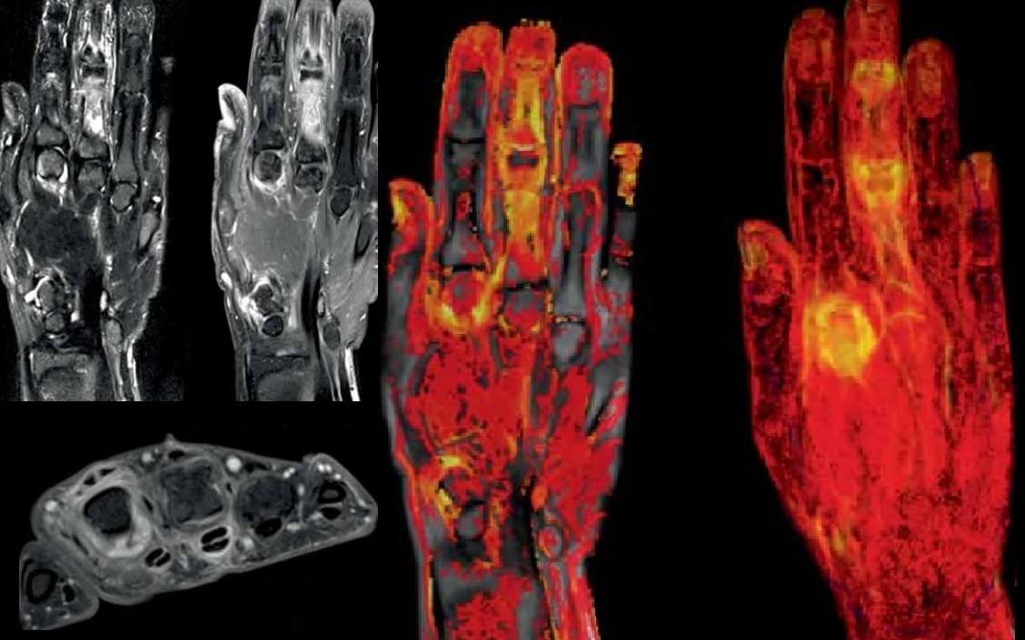
IAG’s team plays an active role in the scientific community and have led the development of novel scoring systems for early assessment of synovial inflammatory changes, quantification of inflammation and quantitative assessment of erosive changes.

Our extensive experience in using radiography, which is a conventional gold standard method for assessment of structural damage in spine and sacroiliac joints. We recommend and assure optimal use of Magnetic resonance Imaging (MRI) that has a number of advantages over plain radiography and ultrasound in assessing disease activity and damage in peripheral joints in PsA. MRI features of PsA include synovitis, dactylitis, tenosynovitis, erosions, bone edema and enthesitis. MRI and anatomic studies have helped researchers develop the concept of synovio-entheseal complex in the pathogenesis of PsA. IAG’s scientific team is actively developing and integrating into the trials novel sequences, including

  • Ultrashort T2 echo for assessment of entheses will help in understanding the similarities and differences between mechanical and inflammatory entheseal changes.
  • Whole-body MRI (WB MRI), with OMERACT as part of novel clinical development.
  • Dynamic contract enhanced MRI for quantitative assessment of inflammation.
  • Quantitative assessment of erosions.

About IAG, Image Analysis Group

IAG is a unique partner to life sciences companies developing new treatment and driving the hope of the up-coming precision medicine. IAG leverages expertise in medical imaging and the power of DYNAMIKA™, our proprietary cloud-based platform, to de-risk clinical development and deliver lifesaving therapies into the hands of patients much sooner. IAG provides early drug efficacy assessments, smart patient recruitment and predictive analysis of advanced treatment manifestations, thus lowering investment risk and accelerating study outcomes.

Acting as imaging Contract Research Organization, IAG’s experts also recognize the significance of a comprehensive approach to asset development. They actively engage in co-development projects with both private and public sectors, demonstrating a commitment to cultivating collaboration and advancing healthcare solutions.

Contact our expert team: imaging.experts@ia-grp.com

READ NEXT CASE STUDY >
Experience: Scoring Systems
  • Psoriatic Arthritis MRI Scoring (PsAMRIS)
  • DEMRIQ, DCE-MRI
  • Exploratory: WB-MRI
Experience: Imaging
  • X-ray
  • Ultrasound
  • DCE-MRI
  • MRI
Publications

Since 2007, over 2000 articles were published to cover scientific discoveries, technology break-throughs and special cases. We list here some critically important papers and abstracts.

Testimonials

Combining our technologies and business advisory services with promising life science companies has yielded spectacular results over the past five years. As a trusted partner to many biotech and pharma companies, IAG’s team is proud to share your words and quotes.
ANKYLOSING SPONDYLITIS

ANKYLOSING SPONDYLITIS

ANKYLOSING SPONDYLITIS

Ankylosing Spondylitis (AS), a form of spondylarthritis (SpA), is a systemic inflammatory rheumatic disease. Although the exact pathogenesis of AS is not yet fully understood, as with rheumatoid arthritis, the swelling, pain, and joint damage that characterize AS are considered to be the result of complex autoimmune and inflammatory processes based on both environmental and genetic factors.

IAG’s team has extensive expertise and track record in supporting the development of novel therapeutics in AS and SpA and helping to take these drugs from early phase all the way to phase 3 and approval. Our expertise is in use of

  • X-ray
  • MRI
  • Dynamic Contrast Enhanced MRI
  • CT

to support disease diagnosis and assessment of treatment response.

IAG’s team plays an active role in the scientific community and have led the development of novel scoring systems for early assessment of synovial inflammatory changes, quantification of inflammation and quantitative assessment of erosive changes. Our extensive experience in using radiography, which is a conventional gold standard method for assessment of structural damage in spine and sacroiliac joints. Our medical science team have worked with drug developers in trials incorporating Computed Tomography (CT), which is a more sensitive method for assessment of structural changes in the spine and sacroiliac joints.

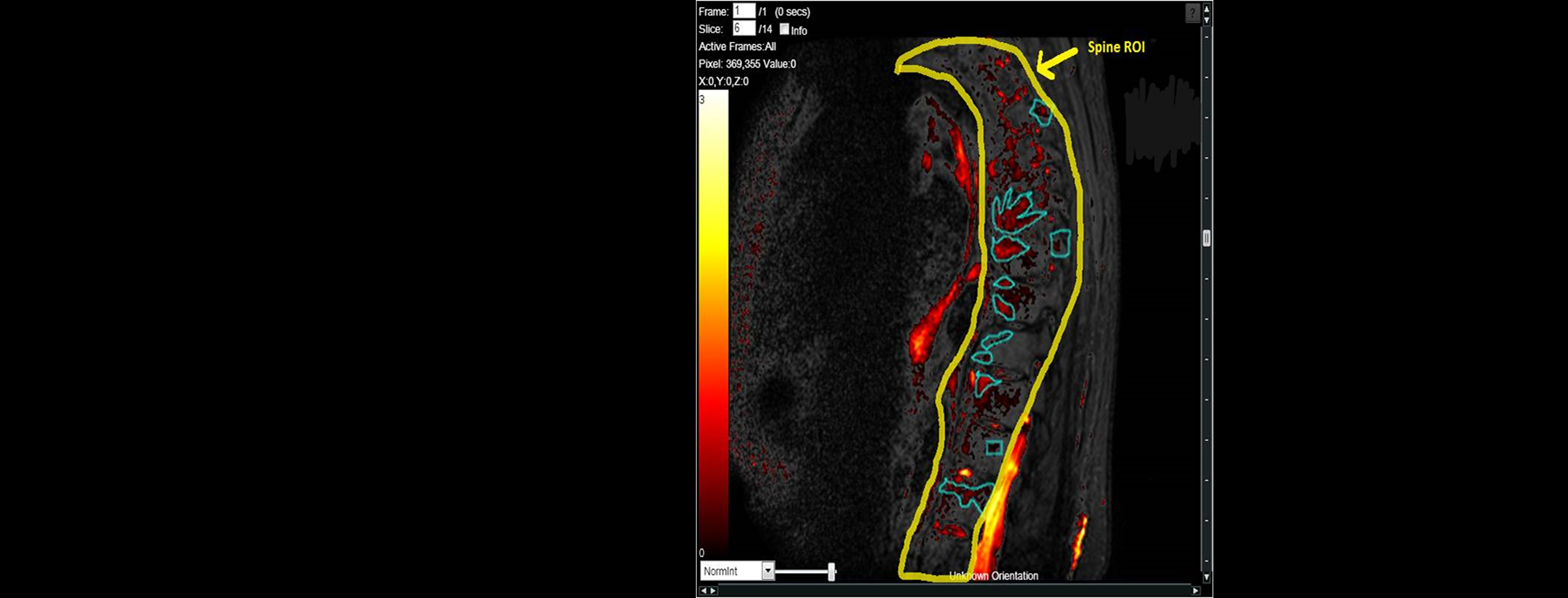
In many trials, early diagnosis of sacroiliitis, the most frequent clinical symptom often accompanied by inflammatory back pain, and other inflammatory lesions of the spine such as spondylitis and spondylodiscitis, can be visualized early by magnetic resonance imaging (MRI), especially using fat saturating STIR or Gd-Enhanced Dynamic MRI sequences. IAG’s team helped to integrate ultrasonography and MRI into the trials for visualization and quantification of peripheral arthritis and enthesitis.


About IAG, Image Analysis Group

IAG is a unique partner to life sciences companies developing new treatment and driving the hope of the up-coming precision medicine. IAG leverages expertise in medical imaging and the power of DYNAMIKA™, our proprietary cloud-based platform, to de-risk clinical development and deliver lifesaving therapies into the hands of patients much sooner. IAG provides early drug efficacy assessments, smart patient recruitment and predictive analysis of advanced treatment manifestations, thus lowering investment risk and accelerating study outcomes.

Acting as imaging Contract Research Organization, IAG’s experts also recognize the significance of a comprehensive approach to asset development. They actively engage in co-development projects with both private and public sectors, demonstrating a commitment to cultivating collaboration and advancing healthcare solutions.

Contact our expert team: imaging.experts@ia-grp.com

READ NEXT CASE STUDY >
Experience: Scoring Systems
  • New York criteria
  • Stoke Ankylosing Spondylitis Spinal Score (SASSS)
  • Bath Ankylosing Spondylitis Radiology Index (BASRI)
  • MRI Spine Score
Experience: Imaging
  • MRI
  • DCE-MRI
  • X-ray
  • CT
  • US
Publications

Since 2007, over 2000 articles were published to cover scientific discoveries, technology break-throughs and special cases. We list here some critically important papers and abstracts.

Testimonials

Combining our technologies and business advisory services with promising life science companies has yielded spectacular results over the past five years. As a trusted partner to many biotech and pharma companies, IAG’s team is proud to share your words and quotes.
JUVENILE INFLAMMATORY ARTHRITIS

JUVENILE INFLAMMATORY ARTHRITIS

JUVENILE INFLAMMATORY ARTHRITIS

Juvenile idiopathic arthritis is the most common autoimmune systemic disease of the connective tissue affecting individuals in the developmental age.  It typically causes joint pain and inflammation in the hands, knees, ankles, elbows and/or wrists and may affect other body parts. IAG’s team supported development of novel treatments in each of the six types of JIA:

  • Oligoarthritis: Affects four or fewer joints, typically the large ones (knees, ankles, elbows).
  • Polyarthritis: Affects five or more joints, often on both sides of the body (both knees, both wrists, etc.). May affect large and small joints.
  • Systemic: Affects the entire body (joints, skin and internal organs).
  • Psoriatic arthritis (PsA): Joint symptoms and a rash.
  • Enthesitis-related: Also known as spondyloarthritis. Affects where the muscles, ligaments or tendons attach to the bone (entheses).
  • Undifferentiated: Symptoms don’t match up perfectly with any of the subtypes, but inflammation is present in one or more joints.

IAG’s team has extensive expertise and track record in using medical imaging to identify and select patients as well as to assess the treatment impact, including use of

  • X-ray
  • MRI
  • US
  • Dynamic Contrast Enhanced MRI
  • Whole-body MRI

Ultrasound assessments involve qualitative assessment of joint cavities, tendon sheaths and bursae for the presence of synovitis, intra-and extraarticular fat tissue to visualize signs of inflammation, hyaline cartilage, cartilaginous epiphysis and subchondral bone to detect cysts and erosions, and ligaments, tendons and their entheses for signs of enthesopathies and tendinopathies.

Quantitative MRI to measure changes in inflammation in peripheral joints, tendon sheaths and bursae, bone marrow assessment and identification of inflammatory lesions in whole-body MRI, particularly when the clinical picture is unclear.

Assessment of MRI of the spine and spinal cord to diagnose synovial joint inflammation, bone marrow oedema and spondylodiscitis as well as to assess their activity, location, and complications (spinal canal stenosis, subluxation, e.g. in the atlantoaxial region).

Assessment of Dynamic Contrast Enhanced MRI to measure the activity of synovitis and confirmation of inflammation in the subchondral bone tissue.

IAG’s team plays an active role in the scientific community and have led the development of novel scoring systems for early assessment of synovial inflammatory changes, quantification of inflammation (bone marrow oedema and synovial) and quantitative assessment of erosive changes. Our expert team and medical team have been working on the assessment of diagnostic and predictive value of single joints quantitative DCE-MRI based imaging biomarkers in JIA as compared to the clinical scores.

About IAG, Image Analysis Group

IAG is a unique partner to life sciences companies developing new treatment and driving the hope of the up-coming precision medicine. IAG leverages expertise in medical imaging and the power of DYNAMIKA™, our proprietary cloud-based platform, to de-risk clinical development and deliver lifesaving therapies into the hands of patients much sooner. IAG provides early drug efficacy assessments, smart patient recruitment and predictive analysis of advanced treatment manifestations, thus lowering investment risk and accelerating study outcomes.

Acting as imaging Contract Research Organization, IAG’s experts also recognize the significance of a comprehensive approach to asset development. They actively engage in co-development projects with both private and public sectors, demonstrating a commitment to cultivating collaboration and advancing healthcare solutions.

Contact our expert team: imaging.experts@ia-grp.com

READ NEXT CASE STUDY >
Experience: Scoring Systems
  • Quantitative Inflammation (DEMRIQ)
  • Juvenile Arthritis MRI Score (JAMRIS)
  • US Doppler
Experience: Imaging
  • MRI
  • Contrast MRI
  • DCE-MRI
  • Ultrasound
  • Radiography
Publications

Since 2007, over 2000 articles were published to cover scientific discoveries, technology break-throughs and special cases. We list here some critically important papers and abstracts.

Testimonials

Combining our technologies and business advisory services with promising life science companies has yielded spectacular results over the past five years. As a trusted partner to many biotech and pharma companies, IAG’s team is proud to share your words and quotes.

OBESITY and SARCOPENIA

OBESITY and SARCOPENIA

BODY COMPOSITION

When conducting an obesity trial to measure body composition, it’s important to choose imaging options that provide accurate and reliable results. Here are some of the best imaging modalities commonly used for this purpose:

  • Dual-Energy X-ray Absorptiometry (DXA): DXA scans are highly accurate and are considered the gold standard for measuring body composition. They can provide information about bone density, fat mass, and lean mass.
  • Computed Tomography (CT) Scan: CT scans can offer detailed information about fat distribution within the body, allowing for precise measurements of visceral and subcutaneous fat.
  • Magnetic Resonance Imaging (MRI): MRI can provide excellent visualization of fat and lean tissue, offering insights into body composition. It’s non-invasive and does not involve radiation.
  • Bioelectrical Impedance Analysis (BIA): While not an imaging modality in the traditional sense, BIA uses electrical impedance to estimate body composition. It’s relatively simple and cost-effective.
  • Ultrasound: Ultrasound imaging can be used to assess the subcutaneous fat thickness and muscle thickness at specific locations, making it useful for localized body composition measurements.
  • Air Displacement Plethysmography (ADP): ADP, commonly measured using the BodPod, calculates body composition based on the principles of air displacement. It’s non-invasive and provides accurate results.
  • Positron Emission Tomography (PET) Scan: PET scans can be used to assess metabolic activity in fat tissue, providing insights into obesity-related metabolic changes.

The choice of imaging modality should depend on factors such as the specific research goals, budget, and accessibility of equipment. It’s often advisable to consult with a medical imaging expert or radiologist to determine the most suitable option for your obesity trial. Additionally, consider ethical and safety aspects when conducting imaging studies involving human participants

About IAG, Image Analysis Group

IAG is a unique partner to life sciences companies developing new treatment and driving the hope of the up-coming precision medicine. IAG leverages expertise in medical imaging and the power of DYNAMIKA™, our proprietary cloud-based platform, to de-risk clinical development and deliver lifesaving therapies into the hands of patients much sooner. IAG provides early drug efficacy assessments, smart patient recruitment and predictive analysis of advanced treatment manifestations, thus lowering investment risk and accelerating study outcomes.

Acting as imaging Contract Research Organization, IAG’s experts also recognize the significance of a comprehensive approach to asset development. They actively engage in co-development projects with both private and public sectors, demonstrating a commitment to cultivating collaboration and advancing healthcare solutions.

Contact our expert team: imaging.experts@ia-grp.com

READ NEXT CASE STUDY >
Experience: Scoring Systems
  • Eligibility and Safety Assessments
  • Body Mass Index (BMI) Score
  • Visceral Adiposity Index (VAI)
  • Fat Mass Index (FMI)
  • Fat-Free Mass Index (FFMI)
  • Total Body Fat Percentage
  • Sarcopenia Index
  • Epicardial Fat Volume Score
  • Liver Fat Score
  • Muscle Quality Score
  • Phase Angle (PhA)
Experience: Imaging
  • MRI
  • DEXA
  • CT
  • PET, PET-CT
  • Ultrasound
  • ADP
  • BIA
Publications

Since 2007, over 2000 articles were published to cover scientific discoveries, technology break-throughs and special cases. We list here some critically important papers and abstracts.

Testimonials

Combining our technologies and business advisory services with promising life science companies has yielded spectacular results over the past five years. As a trusted partner to many biotech and pharma companies, IAG’s team is proud to share your words and quotes.