Advancing Autoimmune Disease Research: The Role of MRI in Idiopathic Inflammatory Myopathies
Advancing Autoimmune Disease Research
Autoimmune diseases remain one of the most complex challenges in modern medicine. These conditions occur when the immune system mistakenly attacks the body’s own tissues, leading to chronic inflammation, organ damage, and significant patient burden. Among these disorders, idiopathic inflammatory myopathies (IIM), a group of autoimmune diseases that affect skeletal muscle, are particularly difficult to diagnose, monitor, and treat.
New research is helping advance the way these diseases are evaluated in clinical trials. A recent study published on PubMed, “The Utility of Muscle Magnetic Resonance Imaging in Idiopathic Inflammatory Myopathies: A Scoping Review,” highlights the growing importance of MRI in assessing inflammatory muscle diseases and tracking treatment response. The research reinforces how imaging biomarkers are becoming essential in autoimmune disease research and clinical trials.
IAG is a proud partner in this research, and this indicates an important shift toward advanced imaging that represents a critical opportunity to improve how autoimmune diseases are studied and treated.
Understanding B-Cell–Mediated Autoimmune Disease
Many autoimmune disorders, including inflammatory myopathies, are driven by abnormalities in B cells, a key component of the immune system responsible for producing antibodies.
In B-cell–mediated autoimmune diseases, these antibodies mistakenly target the body’s own tissues. In inflammatory muscle diseases, the attack leads to progressive muscle inflammation, weakness, and structural damage.
Common forms of inflammatory myopathies include:
- Dermatomyositis
- Polymyositis
- Immune-mediated necrotizing myopathy
“Inflammatory myopathies are complex autoimmune diseases where imaging can help reveal patterns of muscle involvement and support more accurate diagnosis.” – Lisa Stein.
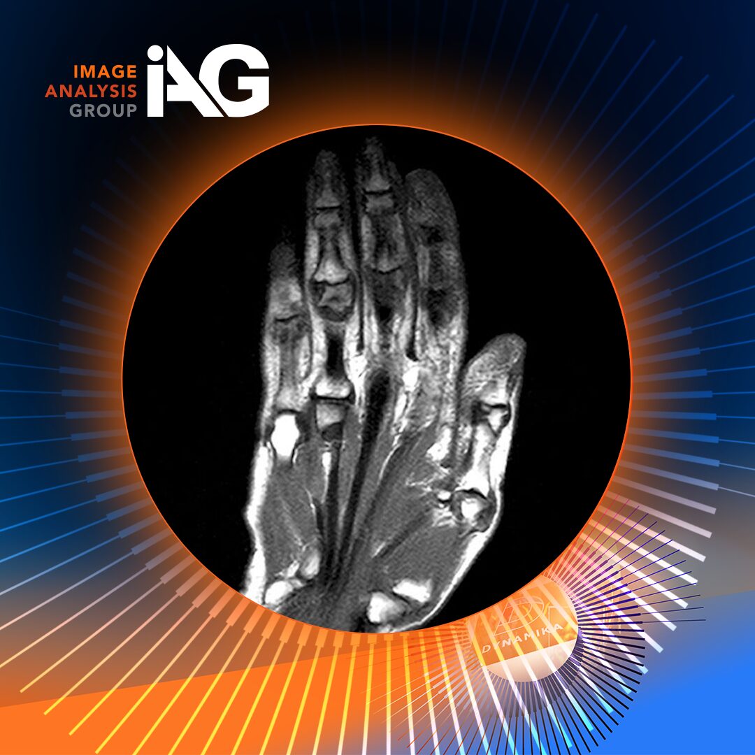
Why MRI Is Critical in Myositis Clinical Trials
Magnetic resonance imaging (MRI) has become one of the most valuable tools for evaluating inflammatory muscle disease.
MRI allows clinicians to visualize key pathological changes, including:
- Muscle inflammation and edema
- Fat infiltration
- Structural muscle damage
In many clinical trials studying inflammatory myopathies, MRI of the thigh muscles is performed multiple times throughout the trial. The thigh is often selected because it contains large muscle groups frequently affected by disease and provides a reliable region for monitoring changes over time.
Repeated imaging during a trial provides critical insight into how inflammation evolves and whether therapies are successfully reducing disease activity.
According to Dr. Olga Kubassova, CEO of Image Analysis Group and a recognized leader in quantitative imaging:
“MRI provides a unique window into the underlying biology of inflammatory muscle disease. When used consistently throughout a clinical trial, it enables researchers to track how muscle inflammation evolves and how patients respond to therapy.”
This longitudinal imaging approach is particularly valuable in autoimmune disease trials, where objective biomarkers are essential for measuring treatment impact.
The Rise of Quantitative Imaging Biomarkers
Traditional MRI interpretation has often relied on visual assessment by radiologists. While valuable, this approach can introduce variability and may not detect subtle changes in tissue structure.
Today, the field is rapidly evolving toward quantitative MRI, where advanced algorithms measure tissue characteristics with high precision.
These measurements may include:
- Muscle fat fraction
- Tissue inflammation levels
- Structural degeneration
As John A Carrino elaborates,
“MRI provides critical information about muscle inflammation and damage that supports both diagnosis and monitoring of inflammatory myopathies.”
At IAG, advanced imaging analytics enable clinical trial teams to convert MRI scans into quantifiable biomarkers, providing more reliable endpoints for evaluating therapies.
Dr. Kubassova explains:
“Quantitative imaging transforms MRI from a diagnostic tool into a powerful biomarker platform for clinical trials. It allows researchers to measure disease progression objectively and detect treatment effects earlier.”
This capability is particularly valuable in complex diseases like inflammatory myopathies, where subtle biological changes may occur before clinical symptoms improve.
CAR-T Therapy: A New Frontier in Autoimmune Disease
While imaging technologies are advancing how diseases are measured, immunology breakthroughs are reshaping how they may be treated.
One of the most promising developments is chimeric antigen receptor T-cell therapy (CAR-T).
Originally developed for cancer treatment, CAR-T therapy works by engineering a patient’s T cells to recognize and eliminate specific target cells. In many cancers, these targets are malignant B cells.
Researchers are now exploring the use of CAR-T therapy in autoimmune diseases driven by pathogenic B cells.
In these cases, CAR-T cells are designed to eliminate the B cells responsible for producing harmful autoantibodies. Early studies suggest that removing these autoreactive B cells may induce long-lasting remission in patients with severe autoimmune disease.
This emerging field, sometimes called CAR-T for autoimmunity, represents a potential paradigm shift in treatment.
For diseases such as inflammatory myopathies, where B-cell activity plays a central role, these therapies could dramatically change the therapeutic landscape.
Why Imaging Partners Matter in Advanced Trials
As innovative therapies such as CAR-T enter clinical trials for autoimmune diseases, high-quality imaging data becomes even more important.
Trials investigating immune-modulating therapies require reliable methods to evaluate:
- Changes in inflammation
- Tissue recovery or degeneration
- Long-term treatment impact
This is where specialized imaging partners like Image Analysis Group play a critical role.
IAG works with global pharmaceutical companies and research institutions to provide:
- Standardized imaging protocols across trial sites
- AI-driven image analysis
- Quantitative imaging biomarkers
- Regulatory-grade imaging endpoints
These capabilities ensure that imaging data collected during trials, such as serial MRI scans of the thigh muscles – can be transformed into meaningful clinical insights.
Dr. Kubassova notes:
“Advanced therapies demand advanced biomarkers. Imaging provides an objective, non-invasive way to evaluate how treatments affect the underlying disease.”
By integrating imaging science with clinical trial design, organizations like IAG help ensure that complex therapies are evaluated with the precision and rigor required for regulatory success.
The Future of Autoimmune Disease Trials
Autoimmune disease research is entering a new era.
Several powerful trends are converging:
- Improved understanding of B-cell–mediated disease mechanisms
- Emerging cell-based therapies such as CAR-T
- Rapid progress in quantitative imaging and AI-driven biomarkers
Together, these advances are reshaping how clinical trials are designed and conducted.
MRI, particularly repeated imaging of the thigh muscles during inflammatory myopathy trials, will remain central to this progress. When combined with advanced analytics, MRI provides a powerful tool for monitoring disease activity and evaluating innovative therapies.
As Dr. Kubassova emphasizes:
“Imaging allows us to see what is happening inside tissues in ways that clinical assessments alone cannot. That insight is critical for accelerating the development of new therapies.”
With partners like Image Analysis Group helping translate imaging data into actionable insights, researchers and pharmaceutical companies are better equipped to develop therapies that address the root causes of autoimmune disease.
For patients living with conditions like inflammatory myopathies, these advances bring renewed hope that future treatments may not only manage symptoms, but fundamentally change the course of disease.
About Image Analysis Group (IAG)
Founded in 2007 and headquartered in London, Image Analysis Group (IAG) is the world’s leading specialist Imaging Clinical Research Organization (iCRO). IAG partners with the world’s top pharmaceutical, biotechnology, and clinical research organizations to design, execute, and deliver imaging-based clinical trial endpoints across oncology, inflammatory disease, neurology, musculoskeletal, cardiovascular, and rare disease indications.
IAG’s proprietary DYNAMIKA™ platform is the most advanced cloud-based clinical trial imaging management system available, integrating AI-driven image analysis, centralized quality control, regulatory-compliant reporting, and real-time trial oversight across global multi-center studies. IAG’s global teams – spanning the United Kingdom, United States, European Union, and India — combine deep medical imaging science, therapeutic area expertise, and regulatory intelligence to accelerate drug development timelines and strengthen the evidentiary quality of imaging endpoints in regulatory submissions.
Partner with IAG to advance quantitative MRI in inflammatory myopathy trials.
Our imaging specialists help pharma and biotech teams turn MRI data into meaningful biomarkers for autoimmune disease research, from study setup through analysis and reporting.
Book a DYNAMIKA™ demo today to explore how an intelligent clinical data platform can streamline operations, improve decision-making, and accelerate your next trial.