OPHTALMOLOGY

OPHTALMOLOGY

Imaging Considerations in Ophtalmology

Diseases related to the human eye range widely based on their localization within the eye. There are eye diseases that are widely prevalent in the population and are manageable (such as macular degeneration and glaucoma), and those that are rare and exceedingly difficult to treat (such as inherited retinal diseases). 

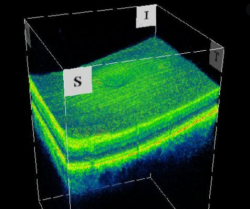
There is a range of highly specialized tests to assess the structural status of the eye that help us detect and monitor these diseases. The management of retinal diseases, such as macular degeneration and retinitis pigmentosa includes a variety of such tests, including ocular coherence tomography (OCT), which uses light waves to take cross-sectional pictures of the retina. Fundus autofluorescence (FAF) and near-infrared reflectance (NIR) provide a density map of lipofuscin and melanin, respectively in the retinal pigment epithelium. These tests are central to the clinical management of these conditions.

Glaucoma, on the other hand, is a common eye disease, and is in fact the most common preventable cause of blindness. It is characterized by an abnormally high intraocular pressure that threatens to damage the optic nerve. It is necessary to be screened with tonometry on routine optometric exams as it may otherwise progress silently and lead to irreversible vision loss. Imaging-based tests for glaucoma include optic nerve head imaging and gonioscopy.

There are several trials being designed and underway that evaluate the safety and efficacy of novel therapeutics for both glaucoma and retinal diseases. These tests, which are critical to the management of these conditions, also serve as primary/secondary endpoints for those trials. These studies need to be meticulously designed to robustly assess these assets for scientific merit, and to deliver safe and effective treatments that are rigorously assessed using these tests. 

At IAG, we are addressing the needs of these trials in the following way:

  • Provide ophthalmologists with high level of expertise in glaucoma and retinal diseases to evaluate/interpret the cases and provide input for drug development strategies.

  • Provide a network of state-of-the art trial sites and testing centers fully capable of executing ophthalmology trials.

  • Provide medical and computational expertise to evaluate structural and functional biomarkers to monitor these diseases.

  • Provide state-of-the-art clinical trials solutions that are nuanced and scientifically optimized.

  • Provide cloud-based infrastructure and software solutions as needed for data exchange between the testing center, trial site and the CRO.

Ophthalmology is an exciting therapeutic area in which the clinical trials need sophisticated solutions. IAG is positioned to meet these needs in cost-effective ways without compromising on benchmarks of scientific methodology and the operational quality.

Reach out to our expert team, as you are designing and planning your trial.

About IAG, Image Analysis Group

IAG is a unique partner to life sciences companies developing new treatment and driving the hope of the up-coming precision medicine. IAG leverages expertise in medical imaging and the power of DYNAMIKA™, our proprietary cloud-based platform, to de-risk clinical development and deliver lifesaving therapies into the hands of patients much sooner. IAG provides early drug efficacy assessments, smart patient recruitment and predictive analysis of advanced treatment manifestations, thus lowering investment risk and accelerating study outcomes.

Acting as imaging Contract Research Organization, IAG’s experts also recognize the significance of a comprehensive approach to asset development. They actively engage in co-development projects with both private and public sectors, demonstrating a commitment to cultivating collaboration and advancing healthcare solutions.

Contact our expert team: imaging.experts@ia-grp.com

READ NEXT CASE STUDY >
Experience: Imaging
  • Ocular Coherence Tomography (OCT)
  • Spectral-domain Optical Coherence Tomography (SD-OCT)
  • Fundus Autofluorescence (FAF)
  • Near-Infrared Fluorescence (NIR)
Publications

Since 2007, over 2000 articles were published to cover scientific discoveries, technology break-throughs and special cases. We list here some critically important papers and abstracts.

Testimonials

Combining our technologies and business advisory services with promising life science companies has yielded spectacular results over the past five years. As a trusted partner to many biotech and pharma companies, IAG’s team is proud to share your words and quotes.

Age-related Macular Degeneration (AMD)

Age-related Macular Degeneration (AMD)

Age-related Macular Degeneration (AMD)

Age-related macular degeneration (AMD) is a serious condition of the eye and responsible for nearly half of the cases of blindness above the age of 60. AMD ranks third, globally, as a cause of blindness after cataract and glaucoma. An estimated 11 million people were affected by AMD in the United States alone, and 170 million worldwide in 2016 [1].

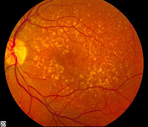
AMD develops on the macula, the central part of retina, where light-sensitive cells are organised with the purpose to capture the image formed inside the eye and pass it dorsally via optic nerves to the brain. The retina, like all body parts, goes through natural wear and tear process with aging. This process manifests itself as toxic yellowish-brown cell debris and deposits called drusens. Gradually, the retina fails to regenerate, reconstitute and renew the parts that are affected by accumulating drusens, which leads to geographical atrophy (cell death) or GA.

Early stage of AMD, also known as “dry” AMD is characterised with drusens and GA and is associated with varying degrees of loss in visual acuity. In the advanced stage, leaky capillaries may form in the mid-layer of the eye, known as choroid layer. Leaking blood and fluid from these capillaries ensues more damage to the macula. This stage is known as “wet” AMD, which, although accounting for a small portion of the AMD cases, is a more serious concern, because it is the cause of 80 to 90% of all legal cases of blindness associated with the disease.

Direct injections of medicine into the eyeball (intravitreal) is the current treatment modality for wet AMD. Type of medications known as anti-angiogenic drugs (anti-VEGF drugs, block vascular endothelial growth factor activity, and cease the formation of new leaky capillaries) may slow down the progression of the disease and enable to partially restore vision.

Clinical imaging techniques have seen expanding applications in the development of effective therapies and tackling the disease, mainly due to the growing number of morphological signs that can assist the early prediction of wet AMD before it causes irreparable damage.

Colour Fundus Photography (CFP) has been a mainstay method for detecting drusens and other morphological changes, by using broad spectrum illumination device. Low contrast and low sensitivity in detecting key biomarkers of AMD, compared to other imaging devices, limits the use CFP.

Fundus Autofluorescence (FAF) imaging, utilizing blue light excitation to acquire retinal images provides sharp contrast and image resolution in detection of atrophic areas on the macula. The pigment retinal epithelium (RPE) and lipofuscin, the “wear-and-tear” pigment of yellow brown, are severely lost in atrophic areas and FAF imaging can provide a strong correlation between structure and function; and intact and degenerating retinal tissue.

While CFP and FAF imaging are clinically practical for grading GA and detecting drusen morphology, both provide limited spatial detection of neovascularization and other wet AMD signalling changes over time. Spectral-domain Optical Coherence Tomography (SD-OCT) can generate in-vivo cross-sectional visualisation of the retina and choroid layer, providing in-depth quantification of time-dependent parameters that have predictive value.

The predictive utility of SD-OCT has been proven in a longitudinal study on 330 AMD eyes in 244 patients, over five years [2]. Algorithms use the patient-derived data to generate a risk score. Being able to predict up to eleven months before discerned progression, investigators have found a formula to successfully quantify AMD associated imaging biomarkers and stratify patients with likelihood of developing wet AMD.

Dynamika, IAG’s imaging data platform, is utilised in clinical development where quantitative imaging biomarkers are pivotal. AMD trials benefit from efficient data management, early efficacy endpoints and increased trial performance transparency.

[1] Pennington K L, DeAngelis M M (2016) “Epidemiology of age-related macular degeneration (AMD): associations with cardiovascular disease phenotypes and lipid factors” Eye and Vision.

[2] Sisternes L, Simon N, Tibshirani R, Leng T, Rubin D L (2014): “Quantitative SDOCT Imaging Biomarkers as Indicators of Age-Related Macular Degeneration Progression” Investigative Ophthalmology and Visual Science Vol. 55 [7093-7103].

READ NEXT CASE STUDY >
Experience: Imaging
  • Colour Fundus Photography (CFP)
  • Fundus Autofluorescence (FAF)
  • Spectral-domain Optical Coherence Tomography (SD-OCT)
Publications

Since 2007, over 2000 articles were published to cover scientific discoveries, technology break-throughs and special cases. We list here some critically important papers and abstracts.

Testimonials

Combining our technologies and business advisory services with promising life science companies has yielded spectacular results over the past five years. As a trusted partner to many biotech and pharma companies, IAG’s team is proud to share your words and quotes.