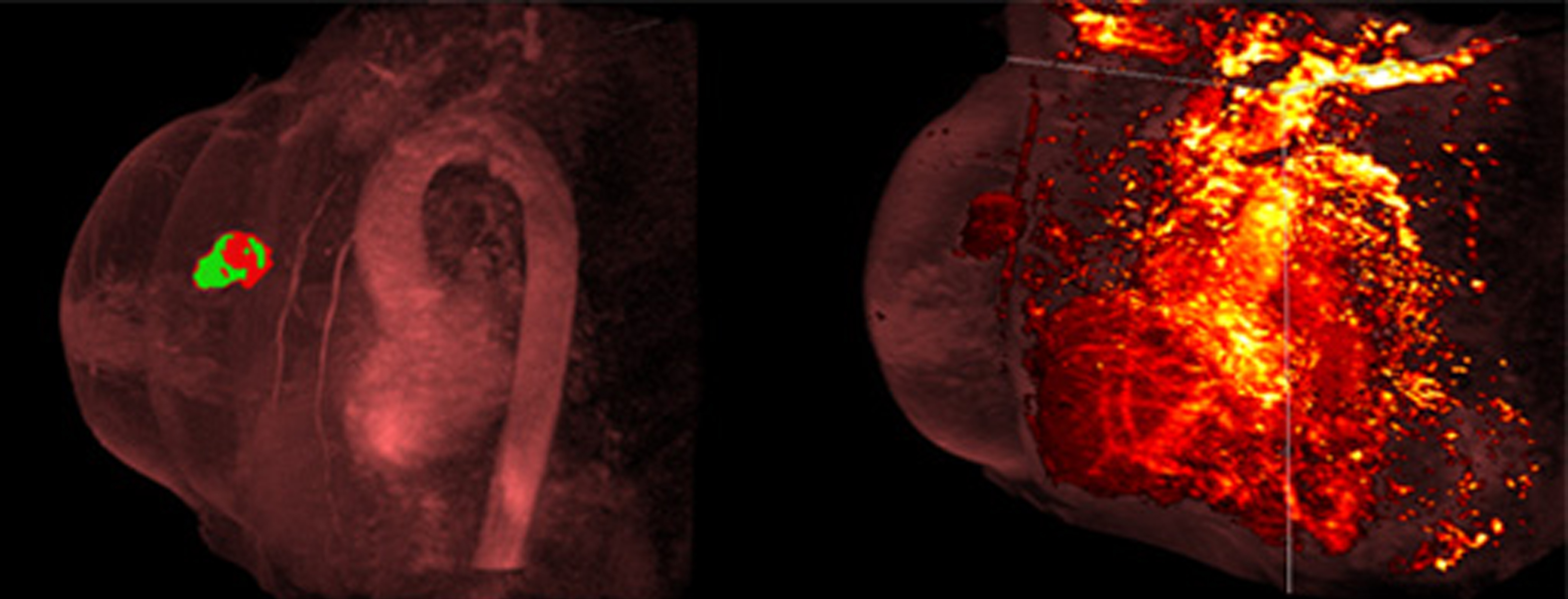

BREAST CANCER
Breast cancer is the most common type of cancer in women worldwide.
Mammography is considered the “gold standard” in the evaluation of the breast lesions from an imaging perspective. Ultrasound examination and Magnetic Resonance maging are being offered as diagnostic techniques and as adjuncts to the pre and postoperative workup. Despite all of these advances, it is still the case that no single imaging modality is capable of identifying and characterising all breast abnormalities and a combined modality approach will continue to be necessary.
Breast MRI is the most sensitive method for detection of breast cancer. Depending on international health regulations, it is either applied for screening of women at high risk for developing breast cancer (e.g. BRCA-1 and BRCA-2 carriers), as an additional diagnostic test in pretherapeutic breast cancer staging, monitoring of primary systemic therapies and for solving problematic diagnostic situations were direct biopsy is not possible.
MRI has exceptional sensitivity for the detection of breast cancer and can depict cancers that are entirely occult on conventional imaging. Reported sensitivities for invasive cancers using dynamic intravenous gadolinium-based contrast agents are consistently greater than 90%.
Dynamic contrast enhanced breast MRI is clinically used to provide volumetric three-dimensional anatomical information and physiologic information that are indicative of increased vascular density and vascular permeability changes associated with angiogenesis.
Perfusion and diffusion imaging techniques may help differentiate between benign and malignant masses. The apparent diffusion coefficient (ADC), a marker of cellularity, is lower in invasive malignancies. Malignant tumours appear to have higher relative blood volumes than normal breast tissue and benign tumours, so perfusion imaging may provide another non-invasive means of tissue characterisation.
Another promising technique in breast cancer diagnose is proton magnetic resonance spectroscopy (MRS). This technique allows for quantitative characterization of total or composite choline concentration that has been shown to be elevated in malignant tumors compared to normal breast tissue. MRS is a nonvasive technique that does not require contrast injection and demonstrates improved sensitivity and specificity when used as an adjunct to breast MRI.
Experience: Scoring Systems
- Breast Imaging, Reporting & Data System (BIRADs)
- UK 5-point scoring system for breast imaging
- RECIST 1.1
Experience: Imaging
- Ultrasound
- Volumetric Breast Ultrasound
- MRI
- DCE-MRI
- DWI-MRI
- MRS
- Mammography
- PET, Positron-Emission Mammography
Publications
Since 2007, over 2000 articles were published to cover scientific discoveries, technology break-throughs and special cases. We list here some critically important papers and abstracts.
Testimonials
Combining our technologies and business advisory services with promising life science companies has yielded spectacular results over the past five years. As a trusted partner to many biotech and pharma companies, IAG’s team is proud to share your words and quotes.

