
CHRONIC RECURRENT MULTIFOCAL OSTEOMYELITIS (CRMO)
Chronic recurrent multifocal osteomyelitis (CRMO) is primarily a diagnosis of exclusion. It is an idiopathic inflammatory disorder of bone seen primarily in children and adolescents.
The imaging evaluation of CRMO should start with radiographic evaluation of the symptomatic sites. If the radiographs are negative in the presence of significant clinical symptoms, further evaluation with MR imaging should be considered to evaluate for marrow edema.
When a diagnosis of CRMO is considered on the basis of clinical findings and initial imaging evaluation, further evaluation of the whole body is suggested to identify multifocal lesions that may be clinically asymptomatic. Whole-body evaluation has traditionally been performed with 99mTc bone scintigraphy, although whole-body MR imaging is being increasingly used for evaluation of multifocal bone lesions.
MR imaging is useful for follow-up of lesions during exacerbation of disease, especially when radiographs show marked sclerosis or hyperostosis. Areas of new activity demonstrate high signal intensity on T2-weighted images and contrast enhancement due to marrow edema, with the surrounding sclerotic bone appearing hypointense on both T1- and T2-weighted images.
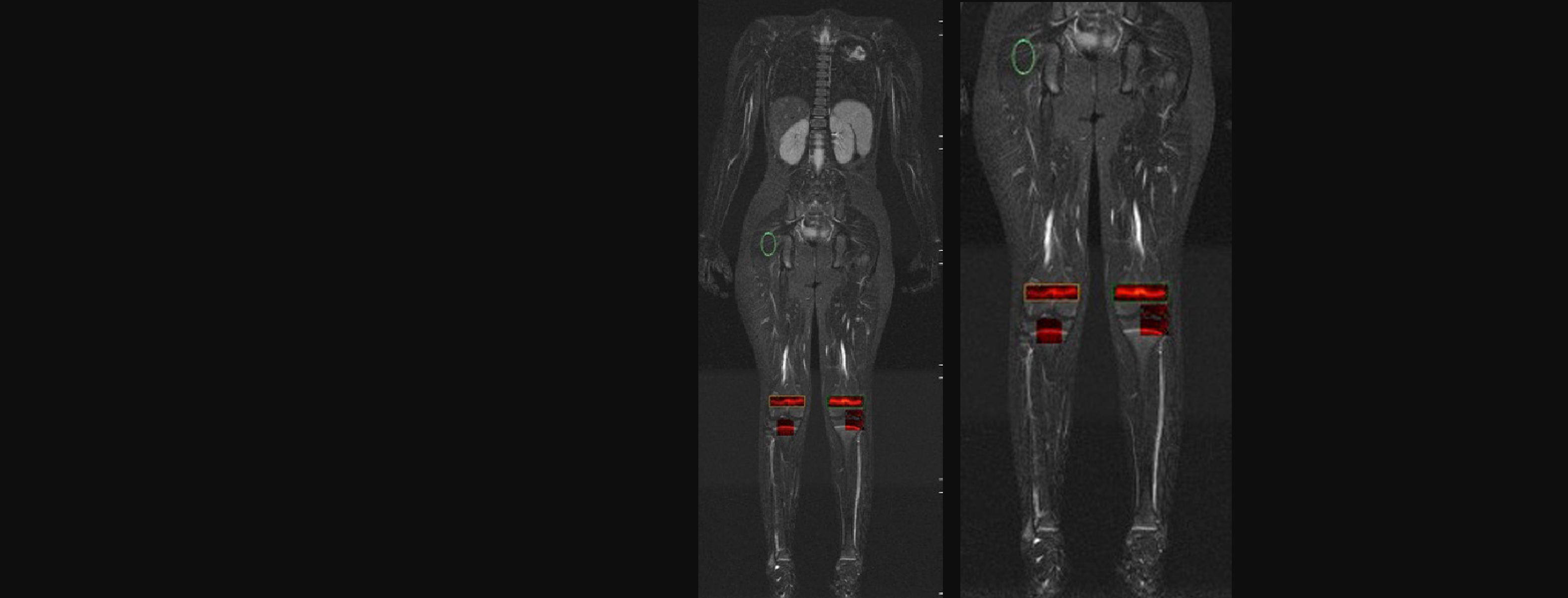
IAG’s team and collaborators presented a novel MRI-based analysis workflow for Chronic Recurrent Multifocal Osteomyelitis (CRMO) at EULAR 2016 and then RSNA 2017. We designed and validated a novel postprocessing scoring method for the quantification of multiple lesions in CRMO patients from static whole body STIR MRI scans, using the normalized intensity (“Norml”) parametric map function, in combination with the region of interest (ROI) tools built into the software Dynamika, a platform for quantitative MRI analysis. The publication discusses the results, where single or multiple lesions seen in whole body MRI scans of CRMO patients were identified and quantified using NormI parametric maps. Quantification parameters for the lesions identified in right femur and right clavicle have been computed by normalising to the baseline in muscle.
Whole body MRI-based workflow for CRMO
Experience: Scoring Systems
- NormI
- DEMRIQ
Experience: Imaging
- MRI T1w
- MRI T2w
- DCE-MRI
- Xray
Publications
Since 2007, over 2000 articles were published to cover scientific discoveries, technology break-throughs and special cases. We list here some critically important papers and abstracts.
Testimonials
Combining our technologies and business advisory services with promising life science companies has yielded spectacular results over the past five years. As a trusted partner to many biotech and pharma companies, IAG’s team is proud to share your words and quotes.

