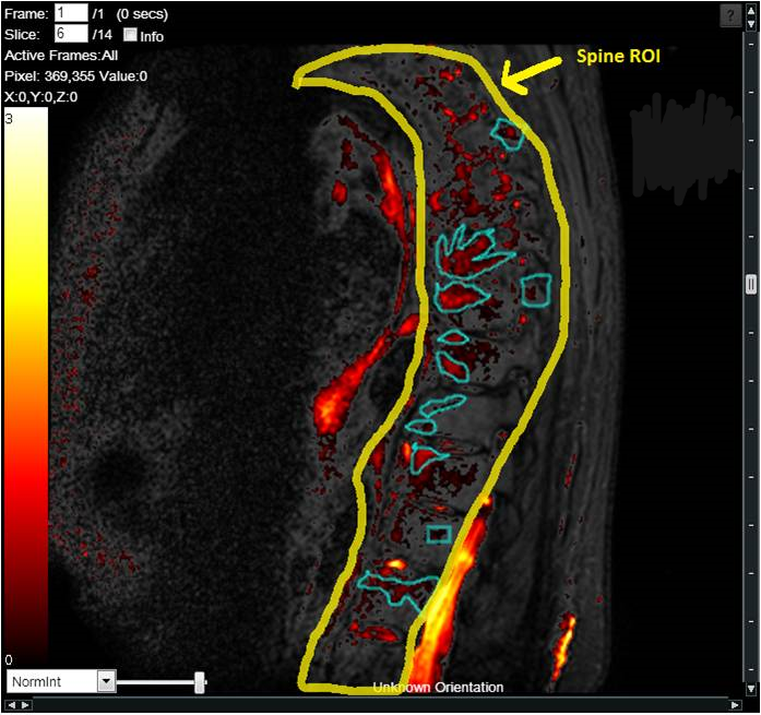

LUMBAR DISC DISEASE / SPINE
Lumbar disk disease may occur when a disc in the low back area of the spine bulges or herniates from between the bony area of the spine. Lumbar disk disease causes lower back pain and leg pain and weakness that is made worse by movement and activity. Degenerative disc disease in the lumbar spine, or lower back, refers to a syndrome in which age-related wear and tear on a spinal disc causes low back pain.
Low back pain (LBP) is the leading cause of disability in developed countries, with the number of people affected worldwide increasing annually.
Back pain is believed to originate from a multitude of lumbar spinal structures including ligaments, facet joints, vertebral periosteum, paravertebral musculature and fascia, blood vessels, annulus fibrosus, and spinal nerve roots.
Therefore, imaging of the lumbar spine has an established role in the evaluation of the LBP patient, and within the last few decades, almost every medical imaging modality has been applied in the evaluation of LBP.
Radiography
Radiography can visualize bony structures and can be used in suspected cases of traumatic, osteoporotic or pathologic vertebral fractures, malalignment, congenital defects, and late stages of inflammatory and infectious diseases. It is an inexpensive and we often recommend it in the initial screening of the spine.
In many of our studies, discography is applied to increase diagnostic accuracy for detecting painful disc levels in LBP with low false-positive rates.
Computed tomography (CT)
Computed tomography (CT) uses a 3D tomographic radiographic technique and is the standard of reference for imaging the vertebral bones, facet joints degeneration, and soft tissue calcification. Tissue contrast on CT is optimal in very dense structures such as bone. Dense soft tissues like tendons, ligaments, and annulus fibrosus can also, to some degree, be differentiated. This makes CT ideal for trauma imaging and fracture characterization in the spine.
We work with a number of CT and DUAL-energy CT (DECT) sites to ensure a new treatment impact has been captured through assessment of the morphology or the chemical composition of the different tissues. Some of the studies include multispectral CT scanners so that our clients have an opportunity to assess change sin bone marrow edema, ligaments, and tendons.
MRI of the lumbar spine
The usual MRI protocols of the lumbar spine include sagittal T1-weighted (T1w) turbo/fast spin echo (TSE/FSE), sagittal and axial T2-weighted (T2w), TSE/FSE images. Adding a sequence in the coronal plane at a later stage has been recommended to allow a better visualization of transitional vertebras, extraforminal disc herniations, extraforaminal nerve roots compression, degenerative vertebral translation, and paravertebral osteophytes.
The presence of fatty tissue in the bone marrow may obscure underlying pathologies on the T2w images without fat saturation. To overcome this issue, spectral fat suppression T2w or fluid sensitive such as the Short Tau Inversion Recovery (STIR) sequences have been used. In STIR sequences, fluid is bright because the contrast is a combination of T1w and T2w images. STIR is the most robust fat-suppression technique in the lumbar spine sensitive for detection of edema and may serve as a screening sequence for many neoplastic, infectious, and traumatic pathologies. However, it is less useful for degenerative conditions because of a high degree of noise and low resolution.
We also design protocols and assess changes using DIXON and DCE-MRI sequences, thus giving more precise information on the pain and inflammation.
IAG’s team has extensive experience in supporting Lower Back Pain, Disc Degeneration ad other Spine disease. Our team plays an active role in the scientific community and have led the development of novel scoring systems and formed strong collaborations with the industry’s Key Opinion Leaders as well as regulatory consultants and agencies.
We bring deep knowledge to help designing a trials, global experienced operations team and fully integrated cloud platform to support a trial that may require use of:
- Xray
- MRI, Dynamic Contrast Enhanced MRI, DIXON, STIR
- CT, PET / CT
About IAG, Image Analysis Group
IAG is a unique partner to life sciences companies developing new treatment and driving the hope of the up-coming precision medicine. IAG leverages expertise in medical imaging and the power of DYNAMIKA™, our proprietary cloud-based platform, to de-risk clinical development and deliver lifesaving therapies into the hands of patients much sooner. IAG provides early drug efficacy assessments, smart patient recruitment and predictive analysis of advanced treatment manifestations, thus lowering investment risk and accelerating study outcomes.
Acting as imaging Contract Research Organization, IAG’s experts also recognize the significance of a comprehensive approach to asset development. They actively engage in co-development projects with both private and public sectors, demonstrating a commitment to cultivating collaboration and advancing healthcare solutions.
Contact our expert team: imaging.experts@ia-grp.com
Experience: Scoring Systems
- MODIC
- Disc morphology
- Inflammation
- Modified Pfirrmann score
- Disc Height
- Spinal Deformity
- DEMRIQ
Experience: Imaging
- X-ray
- MRI
- DCE-MRI
- Ultrasound
- CT
- PET / CT
Publications
Since 2007, over 2000 articles were published to cover scientific discoveries, technology break-throughs and special cases. We list here some critically important papers and abstracts.
Testimonials
Combining our technologies and business advisory services with promising life science companies has yielded spectacular results over the past five years. As a trusted partner to many biotech and pharma companies, IAG’s team is proud to share your words and quotes.

