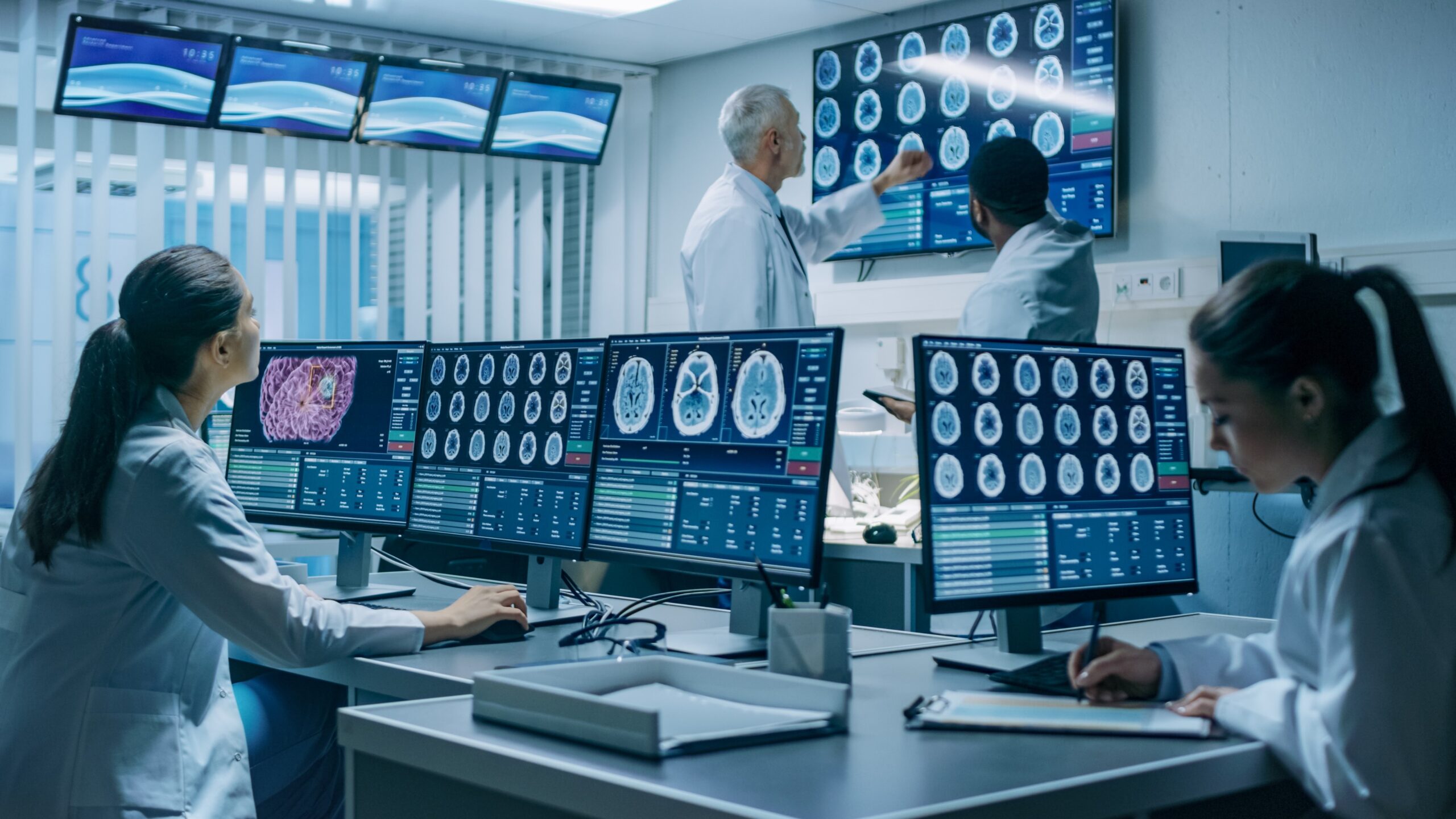

2020 Vision for the Future: Big Data, Predictive Analysis and AI
On November 19 – 22 the 25th Annual Scientific Meeting and Education Day of the Society for Neuro-Oncology (SNO) will be held. The theme this year is 2020 Vision for the Future, and will focus on cutting-edge topics that will shape the future of neuro-oncology, including single-cell sequencing, big data and predictive analytics, biomarkers and liquid biopsy diagnostics, and the role of AI in neuro-oncology research.
This year’s theme emphasizes the global movement in neuro-oncology drug development: a review of standard trial designs, biomarkers and endpoints.
Standard clinical trial designs include overall survival (OS) and progression-free survival (PFS) as primary endpoint.
OS is the gold standard endpoint for large, phase 3 trials in oncology, however, has several challenges. To detect statistically significant differences, larger accruals and longer study durations are needed. This can increase trial budgets and delay reporting of results, which may be difficult to interpret in the context of newer therapies. Additionally, a treatment’s impact on survival may be diluted or confounded by efficacy of salvage therapy— such as radiation therapy for low-grade gliomas—which is often underreported in neuro-oncology trials.
PFS on the other hand hugely relies on the response assessment in neuro-oncology criteria (RANO). To overcome some of the challenges related to OS, PFS might be a better endpoint. Progression free at a prespecified point in time, most often at 6 months (PFS-6), is considered a surrogate marker of OS. However, the relationship and our understanding between response and survival endpoints continues to evolve. For example, PFS and OS failed to correlate in the RTOG 0825 and AvaGlio studies of radiation and temozolomide with or without bevacizumab in newly diagnosed glioblastoma (GBM).
An explanation for these challenges are the nature of novel therapies. Targeted molecular therapies, agents targeting DNA damage response and metabolism, immunotherapies, and viral therapies have complicated mechanism of action and cause complex morphological changes to the tumour that are difficult to capture by RANO’s simple ‘sum of the products’ strategy. At the same time, capturing microenvironmental changes is vital to increase our understanding of the mechanism of action and efficacy of the drug, as well as characterizing responders.
This highlights the need for better suited methodologies to assess response.
Advanced imaging, image analytics and AI/ML can help to overcome RANO’s limitations.
An effective immuno-therapy in GBM will often result in pseudo-progression: an apparent increase in tumour volume, seen on brain scans, which are, in fact, due to tissue necrosis, inflammation and oedema.
This effect poses a challenge to patient retention, management and data interpretation. In addition, due to large variations in the shape of GBM tumor, it is difficult to assess its changes with bi-directional measurements. A novel volumetric semi-automatic segmentation of the true 3D tumour shape can capture the true tumour burden more accurately.

Multiparametric MRI (mpMRI) acquires anatomical and functional MRI sequences and in combination with advanced image processing allow to accurately characterise tumour compartments and measure their longitudinal changes. Highlighted in the image above are oedema tissue in red, contrast-enhancing tissue in purple and necrosis in green. In cases of pseudo-progression and complex tumour microenvironment, RANO easily overestimates the tumour burden, whereas mpMRI often suggests a rather low-moderate genuine tumour burden and more an effect of pseudo-progression that often can be confirmed in a later tumour shrinkage. RANO assessment of the final MRI in the above illustrated case using the ‘sum of the products’ highlights the major shortcoming of the methodology in complex tumor shapes or in small confluent enhancing lesions. RANO suggests stable disease (SD) but the volumetric analysis shows clearly an early response.
IAG’s team is deeply engaged in neuro-oncology drug development and use our proprietary software platform Dynamika for imaging data management and response assessment in clinical trials.
We support our biotech and pharma partners’ clinical trials and help integrating novel analysis methodologies, de-risking and accelerating the development of innovative new treatments for brain cancer.
We will recommend the optimal imaging and help selecting the trial endpoints. Once the trial is designed, IAG’s team will select and train the sites, assist with imaging data collection and review.
Reach out to our expert team, as you are designing and planning your trial.
Engage with our team:
Dr. Diana Dupont-Roettger, Chief Scientific Alliance Officer: https://www.linkedin.com/in/dianaroettger/
About Image Analysis Group (IAG)
IAG, Image Analysis Group is a unique partner to life sciences companies. IAG leverages expertise in medical imaging and the power of Dynamika™ – our proprietary cloud-based platform, to de-risk clinical development and deliver lifesaving therapies into the hands of patients much sooner. IAG provides early drug efficacy assessments, smart patient recruitment and predictive analysis of advanced treatment manifestations, thus lowering investment risk and accelerating study outcomes. IAG bio-partnering takes a broader view on asset development bringing R&D solutions, operational breadth, radiological expertise via risk-sharing financing and partnering models.
Learn more: wp1.ia-grp.com
Reach out: imaging.experts@ia-grp.com
Follow the Company: Linkedin
