ONCOLOGY: Fluorescence imaging

ONCOLOGY: Fluorescence imaging

Fluorescence Imaging

Fluorescence imaging utilizes molecules with fluorescent property – fluorophores – that are excited by specific wavelengths of light, depending on the molecular property.

Subsequently, light is re-emitted with less energy but with longer wavelengths.

The penetration depth of the light depends on the wavelengths.

The near-infrared (NIR) spectrum of 700–1200 nm, the so-called “optical window,” has advantages for in vivo fluorescence imaging as there are less photon scattering, less absorption and less autofluorescence from tissue compared to light in the the visual spectrum. Whereas fluorescence in the visual spectrum can be detected by the human eye, fluorescence in the NIR spectrum requires a digital camera system as light in the NIR spectrum it is not visible to the human eye (Vonk et al, 2021).

Fluorescence guided surgery is an intraoperative medical technique used to generate a real-time fluorescence image to guide the surgical procedure for optimal resection of the tumor (Vonk et al, 2021 and Zheng et al, 2019).


About IAG, Image Analysis Group

IAG is a unique partner to life sciences companies developing new treatment and driving the hope of the up-coming precision medicine. IAG leverages expertise in medical imaging and the power of DYNAMIKA™, our proprietary cloud-based platform, to de-risk clinical development and deliver lifesaving therapies into the hands of patients much sooner. IAG provides early drug efficacy assessments, smart patient recruitment and predictive analysis of advanced treatment manifestations, thus lowering investment risk and accelerating study outcomes.

Acting as imaging Contract Research Organization, IAG’s experts also recognize the significance of a comprehensive approach to asset development. They actively engage in co-development projects with both private and public sectors, demonstrating a commitment to cultivating collaboration and advancing healthcare solutions.

Contact our expert team: imaging.experts@ia-grp.com

READ NEXT CASE STUDY >
Experience: Scoring Systems
  • RANO
  • PET-based Standard Uptake Value (SUV)
  • Volumetric Assessment
Experience: Imaging
  • Fluorescence
  • Anatomical CT / MRI
  • perfusion imaging (DCE-MRI, DSC)
  • diffusion imaging (ADC, DWI, DTI)
  • Multiparametric MRI (mpMRI)
  • PET/CT
  • PET/MRI
  • T-cell Labelling
  • T-Cell Tracing
Publications

Since 2007, over 2000 articles were published to cover scientific discoveries, technology break-throughs and special cases. We list here some critically important papers and abstracts.

Testimonials

Combining our technologies and business advisory services with promising life science companies has yielded spectacular results over the past five years. As a trusted partner to many biotech and pharma companies, IAG’s team is proud to share your words and quotes.

LYMPHOMA

LYMPHOMA

LYMPHOMA

At IAG, Image Analysis Group, we are committed to helping biotech and pharma partners efficiently produce ground-breaking research.

We work behind the scenes, using AI and our proprietary platform DYNAMIKA, to ensure that your study model adheres to the new Lugano guidelines while being cost-effective and acutely aware of other ongoing parallel studies.

In this brief article, we will outline some of the key components of imaging clinical trials of lymphoma that IAG takes into consideration.

With these priorities in mind, rest assured that you can trust IAG every step of the way to help you publish research that will forever change the paradigm of lymphoma treatment.

Currently, the World Health Organization has classified roughly 50 types of lymphoma, with each having numerous subtypes. The following priorities must be statistically analyzed and taken into consideration:

1) Accurate diagnosis and staging. Staging must include whether the lymphoma is limited or advanced, and the degree of tumor burden. The immune status of a patient, for example, such as whether they are immunocompetent or immunocompromised, can significantly alter the imaging uptake response and introduce diagnostic errors.

Many chemotherapy drugs cause cytokine stimulation and inflammatory “flairs,” further muddling MRI, CT, and PET scan findings. Certain tissues heal slower, such as osseous bone, which will show prolonged uptake on contrast PET/CT. At IAG, we ensure none of these errors confound your research.

2) Response to therapy. Each patient must be systematically scanned and cataloged as either complete, partial, stable, progressive, or no response. The Lugano guidelines have recently changed how radiologists measure tumor size and therapy response; our team ensures that all patients are accurately cataloged and tracked to alert you at the first sight of relapse or remission response.

Long term, this develops firm thresholds regarding when to de-escalate or increase therapy to ultimately reduce patient toxicity. At IAG, we also guide our partners in how to choose an appropriate end-point, thus determining therapeutic efficacy.

3) Quality metrics. No imaging modality is 100% accurate. CT and PET scan results are often discordant, with radiologists having known subjective image interpretation and intraobserver variability. The use of IAGs artificial intelligence software eliminates this to offer you standardized reporting and acquisition, with ongoing sensitivity and specificity reports.

4) Record and study the nonmeasurables. Pleural effusions, ascites, cutaneous lesions, metabolic changes, aggressive transformations, and infiltrative developments are just a few of the “nonmeasurables” that science currently has yet to elucidate. At IAG, we aim to one day better characterize these developments, making our partners the leaders in lymphoma research.

Since the Ann Arbor staging system was introduced in 1971, the system has undergone four major overhauls, with oncologists and radiologists alike still calling for a unified guideline and simpler assessment. As a prospective partner, we invite you to join IAG as we radically, forever, change this status quo and cure lymphoma.

About IAG, Image Analysis Group

IAG is a unique partner to life sciences companies developing new treatment and driving the hope of the up-coming precision medicine. IAG leverages expertise in medical imaging and the power of DYNAMIKA™, our proprietary cloud-based platform, to de-risk clinical development and deliver lifesaving therapies into the hands of patients much sooner. IAG provides early drug efficacy assessments, smart patient recruitment and predictive analysis of advanced treatment manifestations, thus lowering investment risk and accelerating study outcomes.

Acting as imaging Contract Research Organization, IAG’s experts also recognize the significance of a comprehensive approach to asset development. They actively engage in co-development projects with both private and public sectors, demonstrating a commitment to cultivating collaboration and advancing healthcare solutions.

Contact our expert team: imaging.experts@ia-grp.com

READ NEXT CASE STUDY >
Experience: Scoring Systems
  • Lugano
  • Olsen
Experience: Imaging
  • CT
  • PET/CT
  • MRI
  • PET
Publications

Since 2007, over 2000 articles were published to cover scientific discoveries, technology break-throughs and special cases. We list here some critically important papers and abstracts.

Testimonials

Combining our technologies and business advisory services with promising life science companies has yielded spectacular results over the past five years. As a trusted partner to many biotech and pharma companies, IAG’s team is proud to share your words and quotes.
LUNG CANCER

LUNG CANCER

LUNG CANCER

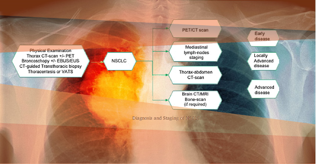
Lung cancer is usually diagnosed in advanced stages with majority of patients already presenting metastatic disease. Only 20 % of NSCLC patients have early stage disease at the time of diagnosis, thus being potentially resectable. The current gold standard is lobectomy with hilar and mediastinal lymph-node sampling or dissection. Curative stereotactic body radiotherapy (SABR) should be offered to patients with stage I NSCLC who have clinical comorbidities or are at very high surgery-related risk, and those who refuse to undergo surgical procedure. Post-operative platinum-based chemotherapy is recommended for all patients with stage II and III surgically resected disease. For patients with locally advanced stage NSCLS, there are different recommended options, including

  • Surgery followed by adjuvant chemotherapy
  • Neoadjuvant chemotherapy followed by surgery
  • Neoadjuvant chemo-radiation followed by surgery

Eligibility for pre-operative or post-operative platinum-doublets with or without radiotherapy should be evaluated in the context of an experienced multidisciplinary team.

The diagnostic evaluation initially focuses on careful physical examination and patient’s history, to identify new symptoms or a significant change in the common respiratory symptoms.

In clinical trials, all patients with suspected lung cancer will undergo a non-invasive chest imaging, including X-rays, CT-scan, and if needed positron emission tomography (PET) with fluorodeoxyglucose (FDG).

Conventional contrast-enhanced chest CT-scan is considered the best exam to detect lung cancer, as it provides detailed information on anatomic location, margins, invasion of surrounding structures or chest wall, and mediastinal lymph nodes involvement.

Use of imaging presents a great opportunity for selecting the right patients and enriching the trial.

IAG offers multiple imaging technique solutions, which are utilized in clinical trials both, while scanning patients for inclusion in the clinical studies, and while assessing the efficacy of ongoing treatments.

  • CT Imaging – Computed Tomography (CT) scan of the chest area is the keystone of lung cancer imaging based on which further management options are decided.
  • SPECT imaging – A single-photon emission computed tomography (SPECT) renders tomographic imaging, which could be a more accurate method for regional valuation by diagnosing radioactivity in all pulmonary lobes, avoiding spatial overlapping.
  • ctDNA testing – Without the risks inherent to biopsy, ctDNA can be attained over time permitting for some serial assessments. Numerous clinical studies have furthermore suggested that ctDNA can be used to identify the occurrence of minimal residual disease (MRD) post-surgical resection in various cancer types, especially lung cancer.
  • EGFR/ALK/ROS/BRAF testing – For targeted therapies, with Mechanism of actions targeting multiple mutations in non-small-cell lung cancer (NSCLC) patients, EGFR/ALK/ROS/BRAF testing imaging techniques are useful to assess the patients for eligibility in the trial as well to assess the treatment efficacy.
  • MRI and ECG – MRI and ECG scans are typically utilized to assess the organ functions, before including patients in the Lung cancer clinical trials.

At IAG, we design and implement protocols for thoracic CT images, assess the need for breath hold and use of intravenous contrast. We support image reconstruction, quality control and reading.

To optimise nodule detection, normally all baseline CTs are read by experienced thoracic radiologists; sometimes located in the local trial centres or at a central site. All discrepancies and adjudication is done by IAG’s imaging specialists.  It is common to ask the readers to identify and record all lung nodules greater than a certain size and diameter. We deploy volumetric measures whenever possible. Our measurements include the volume and maximum intensity projections (MIPs) to aid the detection.

MRI exams are done in addition to CT  when we are trying to assess the drug efficacy in a specific way, such as 1) resolution of the tumour invasion into the chest wall and the mediastinal structures (pancoast tumour); 2) impact on the solid and vascular hilar masses; 3) impact on the diaphragmatic abnormalities or when following-up mediastinal lymphoma.

MR exams are susceptible to motion artefacts such as breathing and require specific software to process. Recently, new applications, such as whole-body MR (WBMR) imaging are being deployed to assess metastatic disease. Diffusion weighted imaging (DWI) is used to assess changes in the tumour cellularity and the integrity of the cellular membrane. The DWI sequence is made susceptible to the differences in water mobility. The motion of water molecules is more restricted in tissues with a high cellular density associated with numerous intact cell membranes (e.g. tumour tissue). This technique can be applied for tumour detection and tumour characterisation and for the monitoring of response to treatment.

PET/CT is a combined imaging technique: CT giving anatomical information and PET giving metabolic information to detect lesions initially not seen on CT and to assess more precise localisation of lesions, delineate them from their surrounding structures and provide more accurate characterisation of a lesion as benign or malignant.

Reach out to our expert team, as you are designing and planning your trial.

About IAG, Image Analysis Group

IAG is a unique partner to life sciences companies developing new treatment and driving the hope of the up-coming precision medicine. IAG leverages expertise in medical imaging and the power of DYNAMIKA™, our proprietary cloud-based platform, to de-risk clinical development and deliver lifesaving therapies into the hands of patients much sooner. IAG provides early drug efficacy assessments, smart patient recruitment and predictive analysis of advanced treatment manifestations, thus lowering investment risk and accelerating study outcomes.

Acting as imaging Contract Research Organization, IAG’s experts also recognize the significance of a comprehensive approach to asset development. They actively engage in co-development projects with both private and public sectors, demonstrating a commitment to cultivating collaboration and advancing healthcare solutions.

Contact our expert team: imaging.experts@ia-grp.com

READ NEXT CASE STUDY >
Experience: Scoring Systems
  • RECIST1.1
  • iRECIST
Experience: Imaging
  • CT
  • PET/CT
  • MRI
  • DWI
  • WBMRI
  • PET
Publications

Since 2007, over 2000 articles were published to cover scientific discoveries, technology break-throughs and special cases. We list here some critically important papers and abstracts.

Testimonials

Combining our technologies and business advisory services with promising life science companies has yielded spectacular results over the past five years. As a trusted partner to many biotech and pharma companies, IAG’s team is proud to share your words and quotes.
Decision Making in Surveillance of High-Grade Gliomas Using Perfusion MRI as Adjunct to Conventional MRI and Artificial Intelligence.

Copyright © 2019 by American Society of Clinical Oncology
Journal of Clinical Oncology. 2019 May;37(15)_suppl doi: 10.1200/JCO.2019.37.15_suppl.2054

Abstract

BACKGROUND:
Surveillance of High-Grade Gliomas (HGGs) remains a major challenge in clinical neurooncology. Histopathological validation is not an option during the course of disease and imaging surveillance suffers from ambiguous features of both disease progression and treatment related changes. This study aimed to differentiate between Pseudoprogression (PsP) and Progressive Disease (PD) using an artificial intelligence (support vector machine – SVM) classification algorithm.
METHODS:
Two groups of patients with histologically proven HGGs were analysed, a group with a single time point DSC perfusion MRI (45 patients) and a group with multiple time point DSC perfusion MRI (19 patients). Both groups included conventional MRI studies prior and after each perfusion MRI. This study design aimed to replicate decision making in clinical practice including multiple previous studies for each patient. SVM training was performed with all available MRI studies for each group and classification was based on different feature datasets from a single or multiple (subtracted features) time points. Classification accuracy comparisons were performed by calculating prediction error rates for different feature datasets and different time point analyses.
RESULTS:
Our results indicate that the addition of multiple time point perfusion MRI combined with structural (conventional with gadolinium-enhanced sequences) MRI features results in optimal classification performance (median error rate: 0.016, lowest value dispersion). Subtracted feature datasets improved classification performance, more prominently when the final and first perfusion studies were included in the analysis. On the contrary, in the single time point group analysis, structural feature-based classification performed best (median error rate: 0.012).
CONCLUSIONS:
Validation of our results with a larger patient cohort may have significant clinical importance in optimising imaging surveillance and clinical decision making for patients with HGG.

Multi-Parametric MRI as Supplement to mRANO Criteria for Response Assessment to MDNA55 in Adults with Recurrent or Progressive Glioblastoma.

Copyright © 2019 by American Society of Clinical Oncology
Journal of Clinical Oncology. 2019 May;37(15)_suppl doi: 10.1200/JCO.2019.37.15_suppl.e13559

Abstract

BACKGROUND:
Modified response assessment in neuro-oncology (mRANO) criteria are widely used in GBM but seem insufficient to capture Pseudoprogression (PsP), which occurs due to extensive inflammatory infiltration, increased vascular permeability, tumor necrosis and edema. mRANO criteria recommend volumetric response evaluation using contrast-enhanced T1 subtraction maps for identifying PsP. Our approach incorporates multi-parametric MRI biomarkers to unravel the true PsP from recurrence or distinguish Pseudo Response (PsR) – following anti-VEGF agents – from delayed (immuno)response.
METHODS:
Multiple time-points MRI (18-24h after convection-enhance delivery of the anti-IL4-R agent MDNA55, then at 30-day intervals) was utilized to determine response. Multi-parametric MRI biomarkers analyzed included (1) 3D-FLAIR-T2-based tumor volume assessment reflecting edema, necrosis and tumor infiltration; (2) 3D-gadolinium-enhanced-based tumor volume estimation reflecting active tumor infiltration, neo-angiogenesis and disrupted blood brain barrier; (3) Dynamic susceptibility contrast-based relative cerebral blood volume (rCBV) measurements for estimation of the vascular tumour properties; and (4) Diffusion weighted imaging – Apparent diffusion coefficient measurements that assess interstitial edema, tumor cellularity and ischemic injury.
RESULTS:
We demonstrate similar imaging phenotypes on conventional FLAIR-T2- and enhanced T1- MR images among different disease states (PsP vs true progression, PsR vs and immuno-response) and describe the perfusion and diffusion MRI biomarkers that improve response staging including PsP masking true progression, PsP masking clinical response, early progression with delayed response, and differentiation between true and PsR. The results are compared with the mRANO-based assessments for concurrence.
CONCLUSIONS:
Incorporating multi-parametric MRI measurements to determine the complex underlying tissue processes enables a better assessment of PsP, PsR and delayed tumour response, and can supplement mRANO-based response assessments in GBM patients undergoing novel immunotherapies.

Early Response Assessment Through Multiparametric MRI Based Endpoints In A Phase II Multicenter Study Evaluating the Efficacy of DPX-Survivac, Intermittent Low Dose Cyclophosphamide (CPA) and Pembrolizumab Combination Study in Subjects with Solid Tumors.

Copyright © 2019 by American Society of Clinical Oncology
Journal of Clinical Oncology. 2019 May;37(15)_suppl doi: 10.1200/JCO.2019.37.15_suppl.e14245

Abstract

BACKGROUND:
Accurate assessment of tumor response to immunotherapy is challenged by pseudoprogression that mimics true progression. Conventional imaging and RECIST assessment do not adequately distinguish between them given their inability to account for changes in the tumor microenvironment. DPX-Survivac is a novel T cell activating therapy that triggers immune responses against tumors expressing survivin and is being studied in this trial in combination with CPA and pembrolizumab in several solid tumors. Multiparametric MRI approaches – dynamic contrast-enhanced MRI and diffusion-weighted imaging MRI are useful for accurate assessment of structural, perfusion and vascular assessment of the lesion and may identify pseudoprogression and compare to the RECIST-based assessment.
METHODS:
The study will enroll up to 226 evaluable subjects in 5 different cohorts: ovarian cancer, HCC, NSCLC, bladder cancer and MSI-H cancer. These subjects will undergo initial imaging 28 days prior to treatment, to be assessed based on RECIST 1.1, and a pre-treatment tumor biopsy for quantitation of survivin and PD-L1 expression and MSI analyses. Treatment for 35 cycles or until disease progression. All patients will have CT images for RECIST 1.1 and iRECIST assessment. A subset of subjects will undergo mpMRI to calculate advanced imaging biomarkers.
RESULTS:
MRI, clinical and patient-reported outcomes will be analyzed.
CONCLUSIONS:
This study will provide important evidence on the utility of mpMRI + CT-based assessment of response to immunotherapy and use it as an adjunct to the CT-based RECIST criteria by providing insight on how tumor lesions are impacted by treatment.

Radiomics in Clinical Trials – The Rationale, Current Practices, and Future Considerations

Radiomics involves deep quantitative analysis of radiological images for structural and/or functional information. – It is a phenomic assessment of disease to understand lesion microstructure, microenvironment and molecular/cellular function. – In oncology, it helps us accurately classify, stratify and prognosticate tumors based on if, how and when they transform, infiltrate, involute or metastasize, – Utilizing radiomics in clinical trials is exploratory, and not an established end-point. – Integrating radiomics in an imaging-based clinical trials involves a streamlined workflow to handle large datasets, robust platforms to accommodate machine learning calculations, and seamless incorporation of derived insights into outcomes matrix.