LIVER CANCER

LIVER CANCER

LIVER CANCER

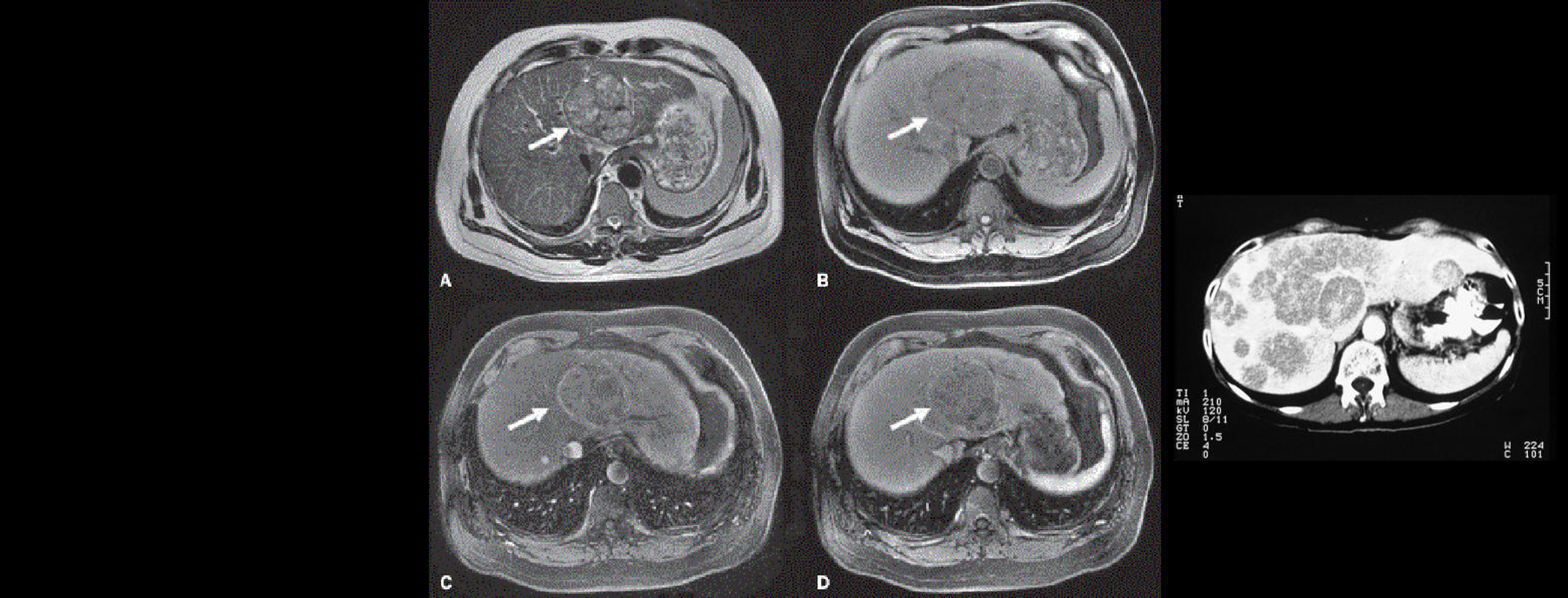
Liver imaging in patients with a history of known or suspected malignancy is important because the liver is a common site of metastatic spread, especially tumours from the colon, lung, pancreas and stomach, and in patients with chronic liver disease who are at risk for developing hepatocellular carcinoma.

The goal of liver imaging in oncologic patients includes liver tumour detection and characterisation.

Liver biopsy is a standard procedure for diagnosing and staging non-alcoholic fatty-liver disease. However, biopsy has a number of disadvantages, including sampling error, intra- and inter-rater variability and poor patient acceptance due to potential complications. Hepatocellular carcinoma (HCC) is the third most common cause of cancer-related death worldwide.

Quantitative imaging is a promising approach for evaluating normal biological or pathogenic processes, as well as response to treatment and intervention without the need for invasive procedures. The field of hepatic MRI is relatively new compared with other organ systems as the complexity of hepatic imaging, including the dual blood supply of the portal vein and hepatic vessels, makes tracer imaging particularly challenging.

HCC commonly develops in the setting of concomitant or previous cirrhotic nodules or hepatic fibrosis, which can complicate classification of suspicious lesions using conventional imaging techniques alone. Depending on the imaging technique used (MRI, CT, US), various quantitative biomarkers can be extracted.

Proton Density Fat Fraction (PDFF)

Chemical-shift imaging is used to separate the liver signal into its fat and water parts.  PDFF is the fraction of MRI-visible protons attributable to fat divided by all protons in the liver attributable to fat and water. This technique acquires GREs  at appropriately spaced echo times. To increase examination accuracy, a low flip angle is used for minimizing T1 bias, along with multiple echoes for correcting T2* effects.

A standard MRI scanner of major manufactures will allow to reproducibly image patients and interpret images. The diagnostic accuracy of MRI-PDFF was validated by several studies, including Idilman et al and Bannas et al, which showed that MRI-based PDFF assessments closely correlated with histology acquired from liver biopsy.

MR Spectroscopy 

MR Spectroscopy, similarly to liver biopsy, is collected from a single region positioned in the liver parenchyma and directly measures the differences in water and fat peaks on a resonance frequency domain. Clearly, the quantification is done only within a particular region which limits the comprehensive understanding of the liver disease. Currently, MRS is not available on all MR scanners, which confines its routine adoption for clinical trials and clinical practice.

MR Electrography 

MR elastography uses a modified phase-contrast pulse sequence to visualize rapidly propagating mechanical shear waves. It is available in MRI scanners by major manufactures and can be acquired simultaneously with  other MRI sequences.  The best known application of MRE is in quantification of hepatic fibrosis.

Dynamic Contract Enhanced MRI

Currently, the use of DCE-MRI in both primary hepatic tumors and metastatic lesions are being investigated, as well as non-oncologic disease processes including hepatocellular fibrosis, cirrhosis, acute liver failure, and post-liver transplant rejection. DCE-MRI and tracer parameters can help differentiate regenerative, benign nodules from more concerning lesions. Sahani et al. reported models that differentiated blood flow and blood volume found differences in moderately or poorly differentiated HCCs. General concepts of differing microvascular structure and selection of appropriate anti-angiogenic therapies are important to maximize treatment response. DCE-MRI may assist in assessment of tumor response even before RECIST criteria can apply; early changes during therapy as demonstrated by perfusion parameters are not often seen on morphologic methods of imaging. Our upcoming publication will detail this further.

Our experience in liver imaging and quantitative biomarkers suggests that while there are multiple challenges in implementing MRI-PDFF, MRS, MRE, DCE-MRI in clinical trials, these techniques are reliable and noninvasive tools to assess hepatic steatosis, fibrosis and microvascular structure.In lights of sophisticated therapeutic developments and high treatment standards, we anticipate that in the next few years  multiparametric MR imaging will become more of a standard, enabling better diagnosis and faster assessments of the treatment response.

References

  • Sommer, W. H. et al. Contrast agents as a biological marker in magnetic resonance imaging of the liver: conventional and new approaches. Abdom. Imaging 37, 164–79 (2012).Eisenhauer, E. A. et al. New response evaluation criteria in solid tumours: revised RECIST guideline (version 1.1). Eur J Cancer45, 228–247. (2009)
  • Yang, X. & Knopp, M. V. Quantifying tumor vascular heterogeneity with dynamic contrast-enhanced magnetic resonance imaging: a review. J Biomed Biotechnol2011, 732848 (2011).
  • Li, S. P. & Padhani, A. R. Tumor response assessments with diffusion and perfusion MRI. J. Magn. Reson. Imaging35, 745–63 (2012).
  • Sourbron, S. P. & Buckley, D. L. Classic models for dynamic contrast-enhanced MRI. NMR Biomed.26(8), 1004–27 (2013).
  • Hylton, N. Dynamic contrast-enhanced magnetic resonance imaging as an imaging biomarker. J. Clin. Oncol.24, 3293–8 (2006).
  • Ferl, G. Z. & Port, R. E. Quantification of antiangiogenic and antivascular drug activity by kinetic analysis of DCE-MRI data. Clin. Pharmacol. Ther.92, 118–24 (2012).
  • Knopp, M., Giesel, F., Marcos, H., von Tengg-Kobligk, H. & Choyke, P. Dynamic contrast-enhanced magnetic resonance imaging in oncology. Top Magn Resnon Imaging12, 301–8. (2001).
  • Padhani, A. & Husband, J. Dynamic contrast-enhanced MRI studies in oncology with an emphasis on quantification, validation and human studies. Clin Radiol56, 607–20 (2001).
  • Padhani, A. Dynamic contrast-enhanced MRI in clinical oncology: current status and future directions. J Magn Reson Imaging16, 407–22 (2002).
  • Tofts, P. Modeling tracer kinetics in dynamic Gd-DTPA MR imaging. J Magn Reson Imaging7, 91–101 (1997).
  • Barral, J. et al. A robust methodology for in vivo T1 mapping. Magn Reson Med64, 1057–67 (2010).
  •  DCE MRI Technical Committee. DCE MRI Quantification Profile, Quantitative Imaging Biomarkers Alliance. Version 1.0. Publicly Reviewed Version. Available from: RSNA.ORG/QIBA
  • O’Connor, J., Jackson, A., Parker, G., Roberts, C. & Jayson, G. Dynamic contrast-enhanced MRI in clinical trials of antivascular therapies. Nat Rev Clin Oncol9, 167–77 (2012).
  • Boesen, M. et al. Automatic Computer Aided Quantification Of Synovitis In Rheumatoid Arthritis Using Dynamic MRI And The Impact Of Movement Correction On Signal To Noise Ratio (SNR) And Region Of Interest (ROI) Analysis [abstract]. Arthritis Rheum60, 773 (2009).
  • Kubassova, O. et al. in Med. Image Comput. Comput. Interv. – MICCAI. Lect. Notes Comput. Sci. Vol. 4792 261–269 (2007).
READ NEXT CASE STUDY >
Experience: Scoring Systems
  • LI-RADS
  • RECIST1.1
  • iRECIST
Experience: Imaging
  • US
  • CT
  • MRI
  • PET
Publications

Since 2007, over 2000 articles were published to cover scientific discoveries, technology break-throughs and special cases. We list here some critically important papers and abstracts.

Testimonials

Combining our technologies and business advisory services with promising life science companies has yielded spectacular results over the past five years. As a trusted partner to many biotech and pharma companies, IAG’s team is proud to share your words and quotes.

PROSTATE CANCER

PROSTATE CANCER

PROSTATE CANCER

Prostate cancer is a disease wherein malignant (cancer) cells form in the tissues of the prostate. This is the second most common cancer diagnosed in men3. Worldwide, prostate cancer is the fourth leading cause of death. At an early stage, prostate cancer may be asymptomatic and often has an indolent course that may only need active surveillance. Most of the prostate cancer cases tend to grow slowly and are low-grade with comparatively limited aggressiveness and low risk.

IAG offers multiple imaging solutions in Prostate cancer, which are currently utilized by the companies in conducting clinical trials, either while scanning patients for inclusion or while assesing the disease progression and efficacy of treatments.

  • Positron Emission Tomography (PET) and MRI – PET/MRI compares the benefits of the metabolic imaging of PET and soft-tissue contrast resolution of MRI. Both these modalities have shown parallel performance in prostate cancer lesion diagnosing by using 18F-choline and 11C as radiotracers.


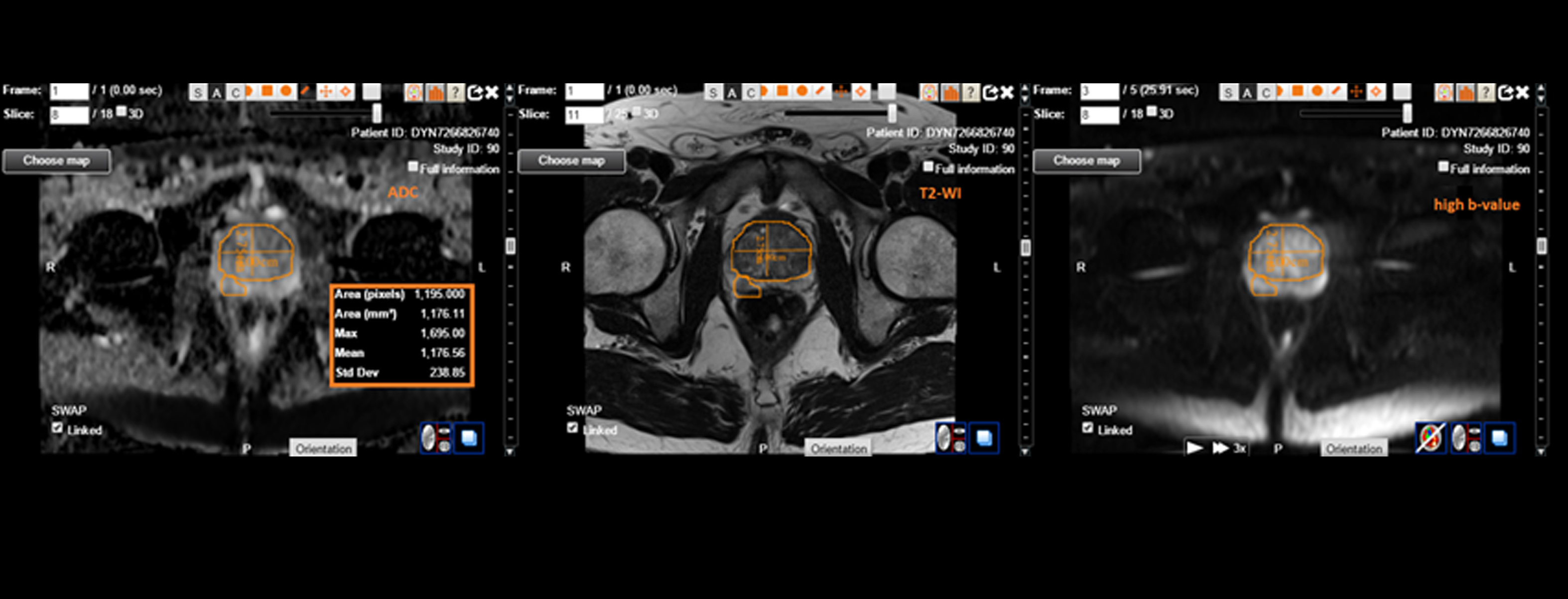
  • Multiparametric-MRI (mpMRI) – multiparametric-MRI (mpMRI) is a recommended MRI technique in prostate cancer (PCa), that comprises high-resolution T2-weighted (T2W) images to understand prostate anatomy and two functional MRI techniques, as well as diffusion-weighted imaging (DWI) to demonstrate dynamic contrast-enhanced MRI (DCE-MRI) and cell density that illustrates vascularity.


  • Bone scan – If prostate cancer spreads to multiple body parts, bones are the first to be impacted. A bone scan can be proven helpful in such cases to demonstrate if the cancer spread has reached the bones.


  • Computed tomography (CT) scan – CT scan helps identify the prostate cancer spread into the nearby lymph nodes and also in cases of recurrence i.e. if it is multiplying into other structures or organs in the patient’s pelvis.


  • PSMA PET – At present, Prostate-specific membrane antigen (PSMA) is considered one of the most efficacious targets for therapy and imaging in the field of nuclear medicine. PSMA is considered to be an outstanding theragnostic agent that delivers the possibility to identify prostate cancer lesions with the help of PET/CT imaging, and later to irradiate metastatic sites with personalized doses by using high-energy alpha or beta particle emitters (radioligand therapy [RLT]).


  • Digital Rectal Examination (DRE) – It is an essential test for the early diagnosis of prostate cancer and is done as part of an annual physical checkup in prostate cancer. Some recent clinical studies show that the combination of PSA and DRE testing is very effective and helpful in the early diagnosis of prostate cancer than each procedure individually.


New-generation diagnostic aids and treatments are evolving and becoming available at a rapid rate that can prove to be very helpful to enhance prognosis in patients with prostate cancer.

IAG, Image Analysis Group offers multiple Prostate Cancer imaging solutions, which are useful while conducting Prostate Cancer clinical trials and which are based on the novel biomarkers emerging in measuring the disease prevalence and disease progression.


We will recommend the optimal imaging and help selecting the trial endpoints. Once the trial is designed, IAG’s team will select and train the sites, assist with imaging data collection and review.

Reach out to our expert team, as you are designing and planning your trial.

About IAG, Image Analysis Group

IAG is a unique partner to life sciences companies developing new treatment and driving the hope of the up-coming precision medicine. IAG leverages expertise in medical imaging and the power of DYNAMIKA™, our proprietary cloud-based platform, to de-risk clinical development and deliver lifesaving therapies into the hands of patients much sooner. IAG provides early drug efficacy assessments, smart patient recruitment and predictive analysis of advanced treatment manifestations, thus lowering investment risk and accelerating study outcomes.

Acting as imaging Contract Research Organization, IAG’s experts also recognize the significance of a comprehensive approach to asset development. They actively engage in co-development projects with both private and public sectors, demonstrating a commitment to cultivating collaboration and advancing healthcare solutions.

Contact our expert team: imaging.experts@ia-grp.com

READ NEXT CASE STUDY >
Experience: Scoring Systems
  • RECIST 1.1 /iRECIST
  • PECIST
  • PET-based Standard Uptake Value (SUV)
  • Volumetric Assessment
  • mpMRI
  • PIRADS
Experience: Imaging
  • CT
  • MRI
  • Perfusion imaging (DCE-MRI, DSC)
  • Diffusion imaging (ADC, DWI, DTI)
  • Multiparametric MRI (mpMRI)
  • PET/CT
  • Multiparametric ultrasound (mpUS)
Publications

Since 2007, over 2000 articles were published to cover scientific discoveries, technology break-throughs and special cases. We list here some critically important papers and abstracts.

Testimonials

Combining our technologies and business advisory services with promising life science companies has yielded spectacular results over the past five years. As a trusted partner to many biotech and pharma companies, IAG’s team is proud to share your words and quotes.
Multi-Parametric MRI As Supplement to mRANO Criteria for Response Assessment to MDNA55 in Adults with Recurrent or Progressive Glioblastoma.

Copyright © 2019 by American Society of Clinical Oncology
Journal of Clinical Oncology May 2019; 37(15)_suppl DOI: 10.1200/JCO.2019.37.15_suppl.e13559

Abstract

BACKGROUND:
Modified response assessment in neuro-oncology (mRANO) criteria are widely used in GBM but seem insufficient to capture Pseudoprogression (PsP), which occurs due to extensive inflammatory infiltration, increased vascular permeability, tumor necrosis and edema. mRANO criteria recommend volumetric response evaluation using contrast-enhanced T1 subtraction maps for identifying PsP. Our approach incorporates multi-parametric MRI biomarkers to unravel the true PsP from recurrence or distinguish Pseudo Response (PsR) – following anti-VEGF agents – from delayed (immuno)response.
METHODS:
Multiple time-points MRI (18-24h after convection-enhance delivery of the anti-IL4-R agent MDNA55, then at 30-day intervals) was utilized to determine response. Multi-parametric MRI biomarkers analyzed included (1) 3D-FLAIR-T2-based tumor volume assessment reflecting edema, necrosis and tumor infiltration; (2) 3D-gadolinium-enhanced-based tumor volume estimation reflecting active tumor infiltration, neo-angiogenesis and disrupted blood brain barrier; (3) Dynamic susceptibility contrast-based relative cerebral blood volume (rCBV) measurements for estimation of the vascular tumour properties; and (4) Diffusion weighted imaging – Apparent diffusion coefficient measurements that assess interstitial edema, tumor cellularity and ischemic injury.
RESULTS:
We demonstrate similar imaging phenotypes on conventional FLAIR-T2- and enhanced T1- MR images among different disease states (PsP vs true progression, PsR vs and immuno-response) and describe the perfusion and diffusion MRI biomarkers that improve response staging including PsP masking true progression, PsP masking clinical response, early progression with delayed response, and differentiation between true and PsR. The results are compared with the mRANO-based assessments for concurrence.
CONCLUSION:
Incorporating multi-parametric MRI measurements to determine the complex underlying tissue processes enables a better assessment of PsP, PsR and delayed tumour response, and can supplement mRANO-based response assessments in GBM patients undergoing novel immunotherapies.

RENAL CELL CARCINOMA (RCC)

RENAL CELL CARCINOMA (RCC)

RENAL CELL CARCINOMA (RCC)

Renal cell carcinoma (RCC) is the most common primary renal malignant neoplasm in adults. It accounts for approximately 90% of renal tumors and 2% of all adult malignancies. The preferred method of imaging renal cell carcinomas is dedicated renal computed tomography (CT). High resolution, reproducibility, reasonable preparation and acquisition time, and acceptable cost allow CT to remain as the primary choice for radiologic imaging.

MRI is an important alternative. The primary limitation of CT is the characterization of hypoattenuation in masses smaller than 8-10 mm, in which pseudo-enhancement may be a problem. In addition, spread to regional lymph nodes in the absence of lymph node enlargement can be missed.

If contrast material cannot be intravenously administered, CT is a poor choice for evaluating renal masses.

MRI is an important alternative to CT as it is much more sensitive. We have carried out the first reported multiparametric MRI (mpMRI) analysis of tumour diffusion and perfusion changes in response to Stereotactic ablative body radiotherapy (SABR) treatment of RCC.

Our latest work ‘Tumour response assessment on multi-parametric MRI after stereotactic ablative body radiotherapy for primary kidney cancer’ was presented as an oral presentation at ICCR 27-30 June 2017, London,  UK.

In the recently completed phase IIb clinical trial in this context (FASTRACK, trial number U1111-1132-5574, https://clinicaltrials.gov/ct2/show/NCT01676428 ), dose delivered was dependent on tumour size with lesions ≤5 cm diameter receiving a single fraction of 26 Gy and larger lesions three fractions of 14 Gy prescribed to the 99% of the target volume. While it has been shown SABR of RCC can achieve high local control rates, variable degrees of morphological tumour shrinkage are typical, with residual masses being common place for sustained periods post-treatment.

Multi-parametric MRI (mpMRI) can be used to investigate radiation induced changes, and in this study, we conduct an exploratory analysis of diffusion and perfusion changes in RCC tumours after SABR using diffusion-weighted (DWI) and dynamic contrast enhanced (DCE) MRI.

Study objectives were to a) establish a methodology for mpMRI evaluation of RCC response to SABR and b) evaluate the potential utility of mpMRI as a biomarker of treatment response.

Results showed that ADC measures and IRE and AUC parametric maps show promise for indicating patient response after SABR. These parameters may provide novel early response biomarkers in a disease for which conventional CT based geometric RECIST response criteria are presently inadequate. Motion of the kidney during mpMRI acquisition and tumour contouring provided challenges to analysis, however we expect our work to implement 3D motion correction to account for kidney movement in DCE MRI will improve data quantification, particularly towards reproducibility of pharmacokinetic maps.


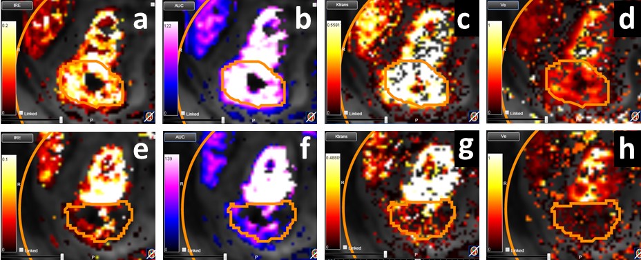
The image above is allustrating DCE MRI maps of a treatment responder pre-SABR (a-d) and post-SABR (e-h) including (left-right) IRE, AUC, Ktrans and Ve with tumour outlined in orange, generated with Dynamika [4]. The corresponding slice between pre-SABR and post-SABR scans.
References

[1]   S. Siva, et al., “A systematic review of stereotactic radiotherapy ablation for primary renal cell carcinoma,” BJU Int., vol. 110, pp. 737–743, 2012.

[2]   D. Pham, et al., “Stereotactic ablative body radiation therapy for primary kidney cancer: a 3-dimensional conformal technique associated with low rates of early toxicity.,” Int. J. Radiat. Oncol. Biol. Phys., vol. 90, no. 5, pp. 1061–8, 2014.

[3]   E. A. Eisenhauer, et al., “New response evaluation criteria in solid tumours: Revised RECIST guideline (version 1.1),” Eur. J. Cancer, vol. 45, no. 2, pp. 228–247, 2009.

[4]   P. S. Tofts, et al., “Estimating kinetic parameters from DCE T1w MRI of a Diffusable Tracer: Standardized Quantities and Symbols,” J. Magn. Reson. imaging, vol. 10, pp. 223–232, 1999.

READ NEXT CASE STUDY >
Experience: Scoring Systems
  • RECIST 1.1
  • irRC (Immune-Related Response Criteria)
  • iRECIST
  • mRECIST
  • PET Response Criteria in Solid Tumors (PERCIST)
  • PET-based Standard Uptake Value (SUV)
  • Volumetric Assessment
  • Dual input function renal perfusion assessment
Experience: Imaging
  • CT
  • DW-MRI
  • DCE-MRI
  • PET / CT
Publications

Since 2007, over 2000 articles were published to cover scientific discoveries, technology break-throughs and special cases. We list here some critically important papers and abstracts.

Testimonials

Combining our technologies and business advisory services with promising life science companies has yielded spectacular results over the past five years. As a trusted partner to many biotech and pharma companies, IAG’s team is proud to share your words and quotes.
HEAD AND NECK CANCER

HEAD AND NECK CANCER

HEAD AND NECK CANCER

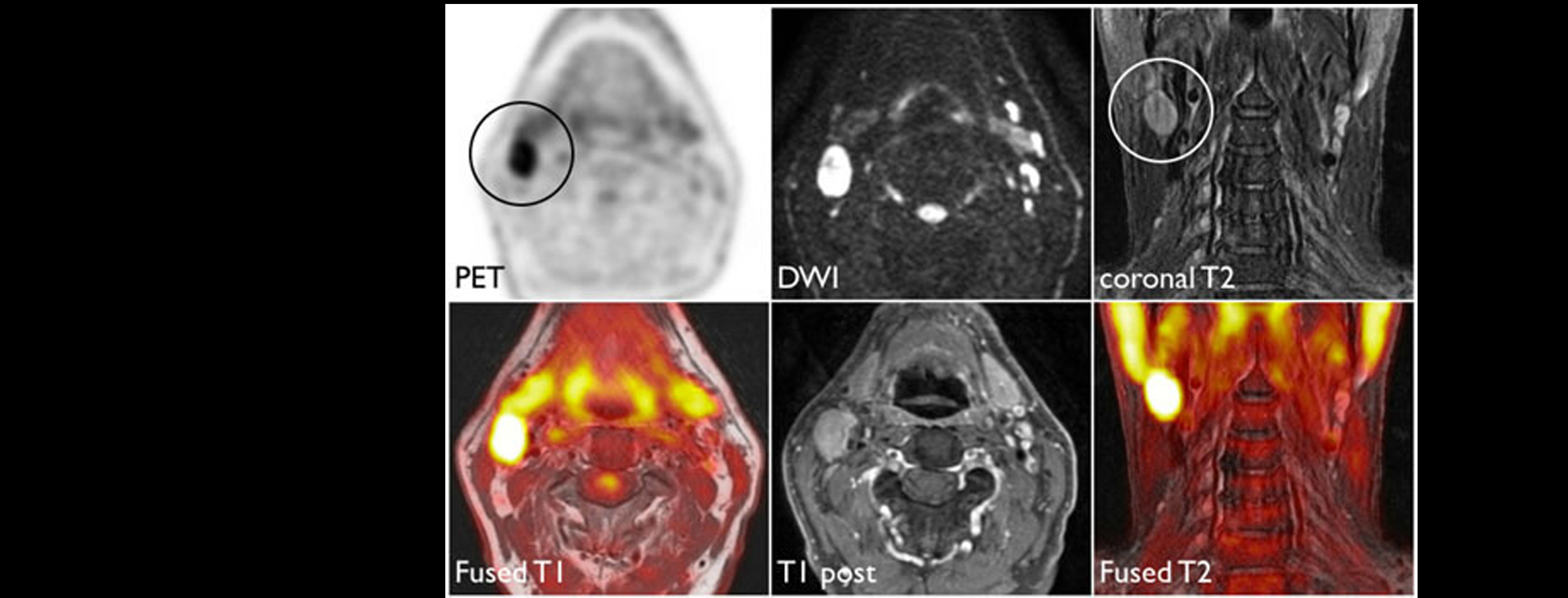
The goals of imaging in head and neck cancer are to establish tumor extent and size, to assess nodal disease, to evaluate for perineural tumor spread, and to distinguish recurrent tumor from post-treatment changes.

MRI is the preferred modality for assessment of nasopharyngeal, sinonasal, and parotid tumors, because of better contrast resolution, high frequency of perineural spread, and less prominent motion artifacts. MRI is the best modality to delineate the extent of intraorbital and intracranial extension of malignant tumors.

Tumors of the oropharynx, larynx, and hypopharynx are frequently primarily imaged with CT, which is less affected by breathing and swallowing artifacts.

MRI is also the initial study of choice for tumors confined to the oral tongue, and possibly also for other oral cavity locations because MRI is superior in detection of tumor spread into the bone marrow.

Positron emission tomography (PET) is very sensitive for metastatic lymph nodes that are at least 8 mm in size and is the technique of choice in dubious cases. For imaging of treated head and neck cancer, PET scans have been found to generally offer higher sensitivity than MRI or CT.

Combined PET/CT may be the modality of choice because it almost completely eliminates the false-positive and false-negative PET findings. Combining PET with MRI has proven to be technically and clinically more challenging than initially expected and, as such, research into the potential clinical role of PET/MRI in comparison with PET/CT, diffusion-weighted MRI (DW MRI) or the combination thereof is still ongoing.

READ NEXT CASE STUDY >
Experience: Scoring Systems
  • RECIST 1.1
  • iRECIST
  • PET-based Standard Uptake Value (SUV)
  • Volumetric Assessment
Experience: Imaging
  • CT
  • Anatomical MRI
  • Perfusion Imaging (DCE-MRI, DSC)
  • Diffusion Imaging (ADC, DWI, DTI)
  • Multiparametric MRI (mpMRI)
  • PET/MRI
  • PET/CT
Publications

Since 2007, over 2000 articles were published to cover scientific discoveries, technology break-throughs and special cases. We list here some critically important papers and abstracts.

Testimonials

Combining our technologies and business advisory services with promising life science companies has yielded spectacular results over the past five years. As a trusted partner to many biotech and pharma companies, IAG’s team is proud to share your words and quotes.

BREAST CANCER

BREAST CANCER

BREAST CANCER

Breast cancer is the most common type of cancer in women worldwide.

Mammography is considered the “gold standard” in the evaluation of the breast lesions from an imaging perspective. Ultrasound examination and Magnetic Resonance maging are being offered as diagnostic techniques and as adjuncts to the pre and postoperative workup. Despite all of these advances, it is still the case that no single imaging modality is capable of identifying and characterising all breast abnormalities and a combined modality approach will continue to be necessary.

Breast MRI is the most sensitive method for detection of breast cancer. Depending on international health regulations, it is either applied for screening of women at high risk for developing breast cancer (e.g. BRCA-1 and BRCA-2 carriers), as an additional diagnostic test in pretherapeutic breast cancer staging, monitoring of primary systemic therapies and for solving problematic diagnostic situations were direct biopsy is not possible.

MRI has exceptional sensitivity for the detection of breast cancer and can depict cancers that are entirely occult on conventional imaging. Reported sensitivities for invasive cancers using dynamic intravenous gadolinium-based contrast agents are consistently greater than 90%.

Dynamic contrast enhanced breast MRI is clinically used to provide volumetric three-dimensional anatomical information and physiologic information that are indicative of increased vascular density and vascular permeability changes associated with angiogenesis.

Perfusion and diffusion imaging techniques may help differentiate between benign and malignant masses. The apparent diffusion coefficient (ADC), a marker of cellularity, is lower in invasive malignancies. Malignant tumours appear to have higher relative blood volumes than normal breast tissue and benign tumours, so perfusion imaging may provide another non-invasive means of tissue characterisation.

Another promising technique in breast cancer diagnose is proton magnetic resonance spectroscopy (MRS). This technique allows for quantitative characterization of total or composite choline concentration that has been shown to be elevated in malignant tumors compared to normal breast tissue. MRS is a nonvasive technique that does not require contrast injection and demonstrates improved sensitivity and specificity when used as an adjunct to breast MRI.

READ NEXT CASE STUDY >
Experience: Scoring Systems
  • Breast Imaging, Reporting & Data System (BIRADs)
  • UK 5-point scoring system for breast imaging
  • RECIST 1.1
Experience: Imaging
  • Ultrasound
  • Volumetric Breast Ultrasound
  • MRI
  • DCE-MRI
  • DWI-MRI
  • MRS
  • Mammography
  • PET, Positron-Emission Mammography
Publications

Since 2007, over 2000 articles were published to cover scientific discoveries, technology break-throughs and special cases. We list here some critically important papers and abstracts.

Testimonials

Combining our technologies and business advisory services with promising life science companies has yielded spectacular results over the past five years. As a trusted partner to many biotech and pharma companies, IAG’s team is proud to share your words and quotes.
ONCOLOGY: ADVANCED IMAGING

ONCOLOGY: ADVANCED IMAGING

ADVANCED ANALYTICS: Oncolytic viral therapy and Immune checkpoint inhibitors

Oncolytic viral therapy and Immune checkpoint inhibitors lead to structural and functional changes within and around the tumor that can be visualized on imaging. These include intra-lesional changes (such as necrosis, hypoxia or associated vascular changes) and perilesional changes (such as edema, hyperemia, leukocyte infiltration).

The immune response triggered by these therapeutic agents is a significant driver of these changes.

The FDA-approved primary endpoint for therapy response to these classes of antineoplastic drugs is the CT-based RECIST 1.1 criteria.

However, our team have worked on and led the development of additional advanced imaging strategies that can be of considerable interest given the information they provide on the pathophysiology and clinical outcomes of solid tumors.

There are several imaging biomarkers derived from standard structural imaging (such as CT) and more sophisticated approaches.

To aid understanding of advanced therapies and to accelerate drug development, at IAG we can help integrating the following imaging methodologies as exploratory endpoints for your trial:

  • Radiomics: This is an automated feature extraction and classification methodology that can analyze sub-visual structural changes detected very early in and around the cancer lesion in response to therapeutic intervention. There are the first order histogram-based and second order texture-analysis based radiomic signatures that can inform on the following:
    • Morphologic changes (spiculation)
    • Cellular density
    • Leukocyte infiltration
    • Vascular changes and resulting hypoxia
    • Necrosis and fibrosis.

These can serve as important biomarkers for therapy response and prognostication/outcomes analysis. They can also be correlated with pathologic data (such as CD3/8 infiltrate count for a more holistic assessment).

  • Molecular imaging: Advanced imaging modalities that evaluate tumors and the immune system at the molecular/cellular level are now translating into human imaging.
    • Reporter gene imaging (bioluminescence, fluorescence imaging) can be used to study the specific activity of oncolytic viruses (biodistribution, infection/transfection, target activity, cell lysis, clearance, therapy response, etc.).
    • ImmunoPET can be very useful for the assessment of cancer cells undergoing lytic changes, and for studying the immune response (both cellular and humoral) to cancer under the influence of immunotherapy.
    • T-cell PET imaging: The cellular immune response as elicited by both these drug groups can be qualitatively and quantitatively assessed with T-cell PET imaging (Visact).

Our work, with leading academic and clinical collaborators is published in a number of articles. Here we provide references to the most relevant articles in the area of advanced analytics.

  • Sun R, Limkin EJ, Vakalopoulou M, Dercle L, Champiat S, Han SR, Verlingue L, Brandao D, Lancia A, Ammari S, Hollebecque A. A radiomics approach to assess tumour-infiltrating CD8 cells and response to anti-PD-1 or anti-PD-L1 immunotherapy: an imaging biomarker, retrospective multicohort study. The Lancet Oncology. 2018 Sep 1;19(9):1180-91.
  • Shaikh F, Franc B, Mulero F. Radiomics as Applied in Precision Medicine. InClinical Nuclear Medicine 2020 (pp. 193-207). Springer, Cham.
  • Shaikh FA, Mulero F, Mohiuddin SA. Molecular Imaging in Genomic Medicine. eLS. 2001 May 30:1-9.
  • Shaikh FA, Kurtys E, Kubassova O, Roettger D. Reporter gene imaging and its role in imaging-based drug development. Drug Discovery Today. 2020 Jan 16.
  • Mayer AT, Natarajan A, Gordon SR, Maute RL, McCracken MN, Ring AM, Weissman IL, Gambhir SS. Practical immuno-PET radiotracer design considerations for human immune checkpoint imaging. Journal of Nuclear Medicine. 2017 Apr 1;58(4):538-46.
  • Tavaré R, Escuin-Ordinas H, Mok S, McCracken MN, Zettlitz KA, Salazar FB, Witte ON, Ribas A, Wu AM. An effective immuno-PET imaging method to monitor CD8-dependent responses to immunotherapy. Cancer research. 2016 Jan 1;76(1):73-82.

Reach out to us to discuss the use of advanced imaging and AI in your trial to support patient stratification, treatment efficacy assessment of patient stratification or tumor signature development, using retrospective historic data.

About IAG, Image Analysis Group

IAG is a unique partner to life sciences companies developing new treatment and driving the hope of the up-coming precision medicine. IAG leverages expertise in medical imaging and the power of DYNAMIKA™, our proprietary cloud-based platform, to de-risk clinical development and deliver lifesaving therapies into the hands of patients much sooner. IAG provides early drug efficacy assessments, smart patient recruitment and predictive analysis of advanced treatment manifestations, thus lowering investment risk and accelerating study outcomes.

Acting as imaging Contract Research Organization, IAG’s experts also recognize the significance of a comprehensive approach to asset development. They actively engage in co-development projects with both private and public sectors, demonstrating a commitment to cultivating collaboration and advancing healthcare solutions.

Contact our expert team: imaging.experts@ia-grp.com

READ NEXT CASE STUDY >
Experience: Scoring Systems
  • RECIST 1.1
  • irRC (Immune-Related Response Criteria)
  • iRECIST
  • mRECIST
  • PET Response Criteria in Solid Tumors (PERCIST)
  • Choi criteria
  • PET-based Standard Uptake Value (SUV)
  • Volumetric Assessment
Experience: Imaging
  • CT
  • anatomical MRI
  • perfusion imaging (DCE-MRI, DSC)
  • diffusion imaging (ADC, DWI, DTI)
  • Multiparametric MRI (mpMRI)
  • PET/CT
  • PET/MRI
  • T-cell Labelling
  • T-Cell Tracing
Publications

Since 2007, over 2000 articles were published to cover scientific discoveries, technology break-throughs and special cases. We list here some critically important papers and abstracts.

Testimonials

Combining our technologies and business advisory services with promising life science companies has yielded spectacular results over the past five years. As a trusted partner to many biotech and pharma companies, IAG’s team is proud to share your words and quotes.
SOLID TUMOURS

SOLID TUMOURS

SOLID TUMOURS

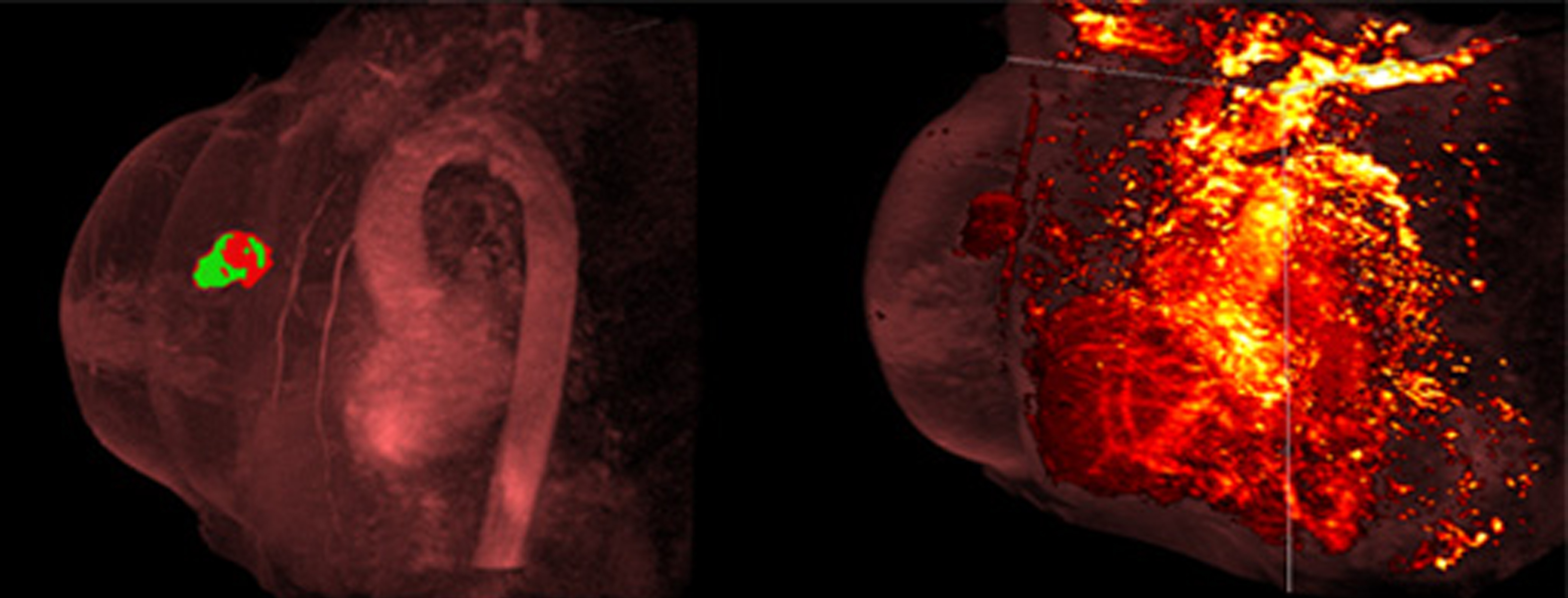
Oncology Imaging is one of IAG’s special areas of therapeutic expertise and focus.  Oncology studies comprise almost one third of IAG’s clinical development experience. Currently IAG is engaged in trials with umbrella, adaptive and basket trial designs. IAG is providing full imaging service and image review in early and late phase trials, bringing our world-wide site network, operational expertise and technology platform DYNAMIKA to manage, collect, hold and score the imaging data.

A large proportion of our studies are the early clinical and phase II studies in solid tumours, lymphomas and brain tumours.

IAG’s radiology and analytics teams have hands-on experience with state-of-the-art imaging scoring systems and novel AI-driven methodologies. We have worked with studies requiring MRI, CT, X-ray, PET, immunoPET, SPECT, US, histology, and fluoroscopy imaging. Specifically, our team is board certified and experienced in:

  • RECIST1.1, iRECIST, mRECIST, irRC, irRECIST,
  • PERCIST and EORTC criteria for PET response,
  • CHOI criteria for response to tumours,
  • Macdonald, RANO, iRANO, mRANO and RAPNO for Neuro-Oncology,
  • PIRADS and PCWG3 criteria for Prostate cancer,
  • Cheson, Lugano and RECIL for lymphoma,
  • Olsen criteria; IWCLL for CLL; MDA for bone metastases.

IAG’s team is an active scientific contributor and our work involved development of quantitative methodologies for

  • Pseudo progression measurements
  • Radiomic Analyses
  • Quantitative assessment of the lesions (texture, shape, heterogeneity)
  • Use of advanced imaging to assess the perfusion, hypoxia, cellular density of the tumours
  • Macrophage Analysis
  • Automated quantitation of total tumour burden and volumetric analysis
  • Use of PET / SPECT novel agents for immune trafficking and receptor imaging

Our experts often use advanced functional imaging to aid early assessment of treatment response in oncology trials. These include:

  • Perfusion measurements
  • Diffusion and cellular density measurements
  • Metabolic and proliferation assessments

We have experience with radiotherapies and advanced therapies, including

  • Cellular therapy
  • Viral therapy
  • Intra tumoral injection
  • Vaccines
  • Combination studies
  • Ablative therapy, radiation therapy
  • Immune system stimulators
  • Radiotherapies

IAG’s in-house radiologists and experts in Artificial Intelligence, Machine Learning and Big Data analytics can assist with all aspects of the imaging trial design, imaging data collection, site feasibility and training, image review and quantitative analysis.

We are proud to support you and bring real precision into the imaging trial, including understanding of pseudo progression and other complex manifestations of the disease in response to treatment.

Our partnerships with oncology focused companies have been published and are available to view on our website at ia-grp.com/trial-solutions/bio-partnering/

Reach out to our expert team, as you are designing and planning your trial.

About IAG, Image Analysis Group

IAG is a unique partner to life sciences companies developing new treatment and driving the hope of the up-coming precision medicine. IAG leverages expertise in medical imaging and the power of DYNAMIKA™, our proprietary cloud-based platform, to de-risk clinical development and deliver lifesaving therapies into the hands of patients much sooner. IAG provides early drug efficacy assessments, smart patient recruitment and predictive analysis of advanced treatment manifestations, thus lowering investment risk and accelerating study outcomes.

Acting as imaging Contract Research Organization, IAG’s experts also recognize the significance of a comprehensive approach to asset development. They actively engage in co-development projects with both private and public sectors, demonstrating a commitment to cultivating collaboration and advancing healthcare solutions.

Contact our expert team: imaging.experts@ia-grp.com

READ NEXT CASE STUDY >
Experience: Scoring Systems
  • RECIST1.1, iRECIST, mRECIST, irRC, irRECIST,
  • Volumetric,
  • Radiomics and Multi-Omics,
  • PERCIST and EORTC criteria for PET response,
  • CHOI criteria for response to tumours,
  • Macdonald, RANO, iRANO, mRANO and RAPNO for Neuro-Oncology,
  • PIRADS and PCWG3 criteria for Prostate cancer,
  • Cheson, Lugano and RECIL for lymphoma,
  • Olsen criteria; IWCLL for CLL; MDA for bone metastases
Experience: Imaging
  • CT
  • MRI
  • PET/CT
  • PET/MRI
  • Perfusion imaging (DCE-MRI, DSC)
  • Diffusion imaging (ADC, DWI, DTI)
  • Multiparametric MRI (mpMRI)
  • T-cell Labelling
  • T-Cell Tracing
  • MRI Spectroscopy
  • Breast-based MRI, digital mammography, US, tomosynthesis
  • US
  • Angiography
Publications

Since 2007, over 2000 articles were published to cover scientific discoveries, technology break-throughs and special cases. We list here some critically important papers and abstracts.

Testimonials

Combining our technologies and business advisory services with promising life science companies has yielded spectacular results over the past five years. As a trusted partner to many biotech and pharma companies, IAG’s team is proud to share your words and quotes.

NEURO-ONCOLOGY

NEURO-ONCOLOGY

NEURO-ONCOLOGY

High-grade gliomas (HGG), is the most common malignant brain tumor, with an incidence of <10 in 100.000 people (Ostrom et al, 2019). The overall prognosis is poor with a median survival of approximately 15 months with optimal treatment offered in clinical trials (Stupp et al 2005). The reported 5 year survival rate varies in the literature, depending on clinical trials or population-based studies but is usually considered less than 10% (Ostrom et al, 2019, Dressler et al 2019, Narita al, 2015, Fuentes Raspall et al, 2017, Poon et al 2019).

The complex intra-tumoral cellular heterogeneity and the intricate pattern of involved genes and underlying mutations makes glioblastoma one of the most difficult cancers to treat (Hatoum et al., 2019; Sottoriva et al, 2013, Shergalis et al., 2018). HGG invades surrounding brain tissue and exerts damaging impact on brain function. Despite considerable attempts to improve surgery, radiation, and medical treatments, no sufficient efficient therapy is available, and the unmet medical need is apparent.

Gliomas are divided into four grades according to the WHO classification with grade I being circumscribed and usually benign if sucessfully resected and grade IV, glioblastoma, being highly malignant (Louis et al, 2016).
HGG can either evolve spontaneously as de novo (primary) accounting for more than 80% of the subjects or develop from lower grade gliomas (secondary GBM) (Schouwer et al. 2017).

In HGG upregulated uPAR expression is observed (Yamamoto et al., 1994, Persson et al., 2016) and there is a significant correlation between increased uPAR expression and increased tumor grade (Salajegheh et al., 2005).

Neuro-Oncology is IAG’s area of expertise and clinical research focus. We work with biotech and pharma companies, who at the forefront of discovery and development of new treatments for brain and central nervous system tumours. IAG’s team have extensive expertise in designing and delivering trials aimed at advancing the understanding, diagnosis, and treatment of brain tumours. Areas of focus include:

  • Immunotherapy
  • Cancer genetics/genotyping
  • Targeted therapies/precision cancer medicine
  • Radiation and chemotherapy

Our scientific advisory board members and leading academic collaborators have made important inroads in basic and clinical research to better understand the biology that underlies these tumours and identify and test new therapies. IAG is involved in several global consortiums and have led the development of novel imaging endpoints in neuro-oncology.

We provide our biotech and pharma partners with extensive expertise in management and treatment development for benign and malignant brain tumors including glioma, ependymomas, glioblastoma multiforme (GBM), medulloblastomas and other adult and paediatric brain cancers.

IAG brings years of oncology, neuro-radiology and quantitative medical imaging expertise to help understanding and assessment of

  • highly complex nature of the tumour,
  • heterogenous tumour microenvironment,
  • pseudo-progression,
  • tumour volume and size changes,
  • Cellular level response.

Any successful therapy must target the inherent tumour heterogeneity. This leads to individually complex structural responses in the tumour microenvironment.

IAG’s team has deep understanding of challenges associated with design and execution of neuro-oncology trials.

We understand that optimal clinical trial design is crucial. Chosen imaging modality and associated image analysis will help to prove the efficacy of the therapy. These must show the functional and anatomical structure of the tumour and help addressing the treatment induced changes quantitatively and as early in the process as possible.

We will recommend the optimal imaging and help selecting the trial endpoints. It is often the case that conventional imaging methods such as anatomical MRI in combination with RANO are adequate, but they also may be misdealing. Once the trial is designed, IAG’s team will select and train the sites, assist with imaging data collection and review.

Reach out to our expert team, as you are designing and planning your trial.

About IAG, Image Analysis Group

IAG is a unique partner to life sciences companies developing new treatment and driving the hope of the up-coming precision medicine. IAG leverages expertise in medical imaging and the power of DYNAMIKA™, our proprietary cloud-based platform, to de-risk clinical development and deliver lifesaving therapies into the hands of patients much sooner. IAG provides early drug efficacy assessments, smart patient recruitment and predictive analysis of advanced treatment manifestations, thus lowering investment risk and accelerating study outcomes.

Acting as imaging Contract Research Organization, IAG’s experts also recognize the significance of a comprehensive approach to asset development. They actively engage in co-development projects with both private and public sectors, demonstrating a commitment to cultivating collaboration and advancing healthcare solutions.

Contact our expert team: imaging.experts@ia-grp.com

READ NEXT CASE STUDY >
Experience: Scoring Systems
  • RANO, iRANO, mRANO
  • Volumetric Assessment
  • Perfusion-based Tumor Vascularity Biomarkers
  • Diffusion-based Cellular Density Biomarkers
  • Macdonald criteria
  • Machine learning techniques for grading of GBM
Experience: Imaging
  • Anatomical MRI (T1, T2, FLAIR)
  • Perfusion Imaging (DCE-MRI, DSC)
  • Diffusion Imaging (ADC, DWI, DTI)
  • Multiparametric MRI (mpMRI)
  • Contrast enhanced MRI (Gadolinium, Ferumoxytol)
  • PET/CT
  • PET/MRI
  • Fluroscence Imaging
Publications

Since 2007, over 2000 articles were published to cover scientific discoveries, technology break-throughs and special cases. We list here some critically important papers and abstracts.

Testimonials

Combining our technologies and business advisory services with promising life science companies has yielded spectacular results over the past five years. As a trusted partner to many biotech and pharma companies, IAG’s team is proud to share your words and quotes.