SCLERODERMA

SCLERODERMA

SCLERODERMA

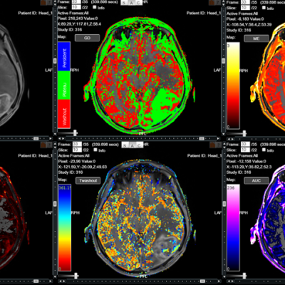
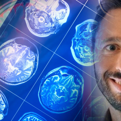
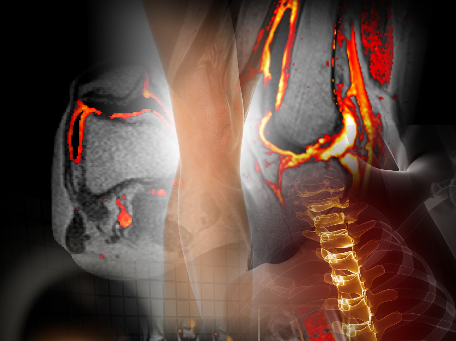
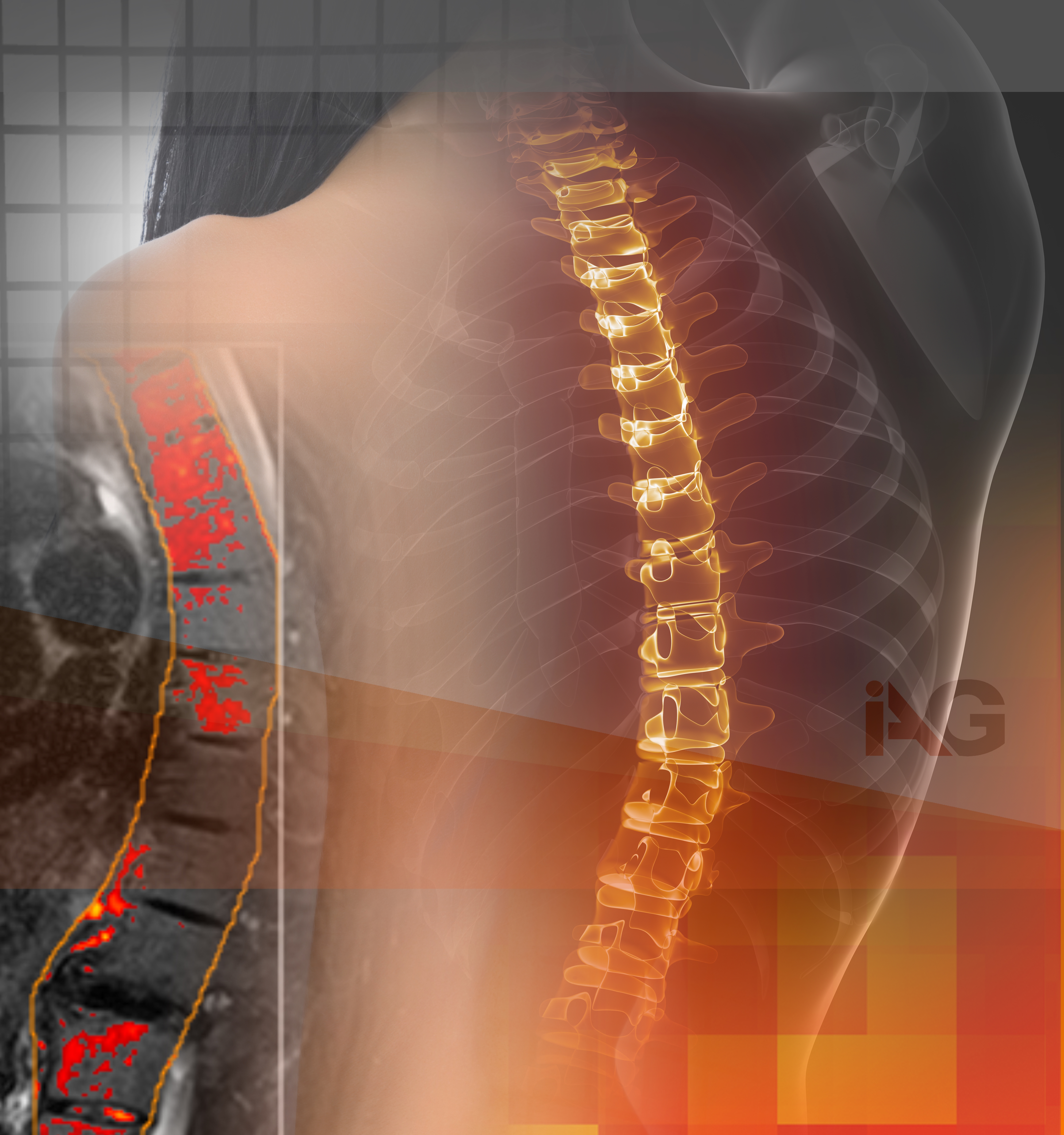
Scleroderma, a rare and often perplexing autoimmune disorder, presents numerous challenges for both patients and drug development organizations. One key tool in unravelling the mysteries of this condition is medical imaging. From diagnosing scleroderma to monitoring disease progression and assessing treatment effectiveness, various imaging modalities play a crucial role in the care of individuals affected by this disease.

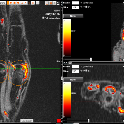
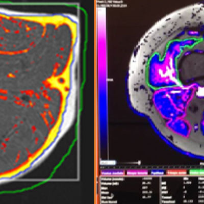
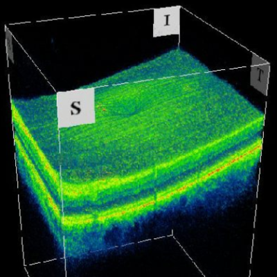
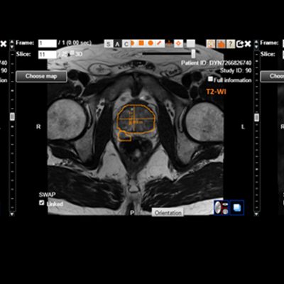
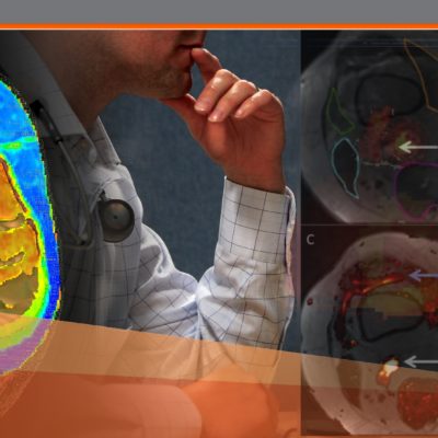
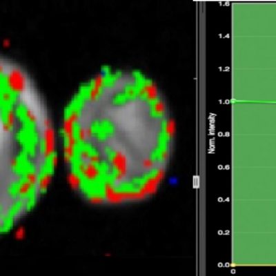
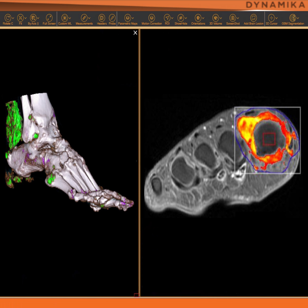
IAG team helps pharmaceutical companies to use medical imaging and associated digital endpoints to support diagnosis / screening, assessment of organ involvement, disease progression monitoring and drug efficacy assessment using MRI, US, Xray, CT and other available imaging modalities.

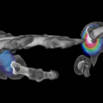
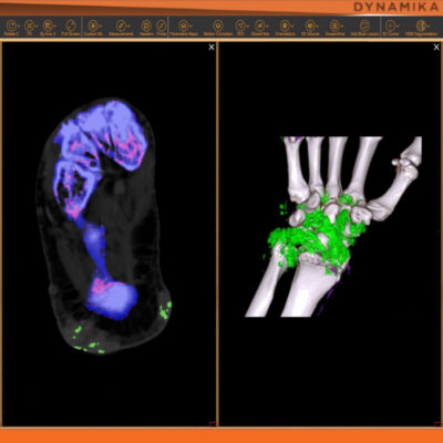
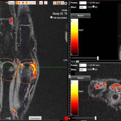
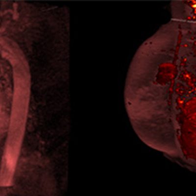
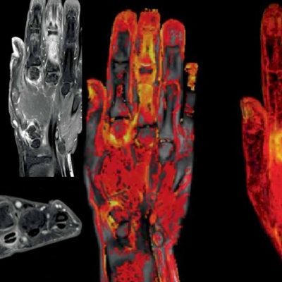
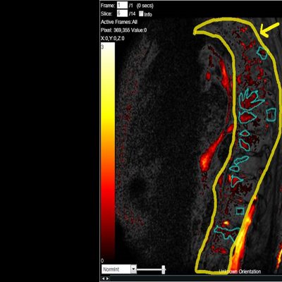
IAG and University of Leeds, UK have partnered to develop a novel Digital Arteries Volume Index, DAVIX, which was used in drug development trials since 2010. DAVIX© has been validated as a predictor of the onset of Digital Ulcers in Systemic Sclerosis patients. This innovative methodology is based on the Time-of-Flight Magnetic Resonance Imaging (MRI) angiography and allows to quantitatively assess digital arterial blood flow.

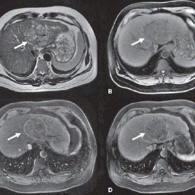
  1. Screening: Scleroderma’s hallmark symptom is skin thickening, but it can also affect internal organs, making early and accurate diagnosis critical. Imaging, such as high-resolution ultrasound and magnetic resonance imaging (MRI), is instrumental in detecting abnormalities within affected tissues and organs. These techniques help clinicians visualize the extent and location of fibrosis, a key feature of scleroderma, and can aid in differentiating the disease from other conditions with similar symptoms.
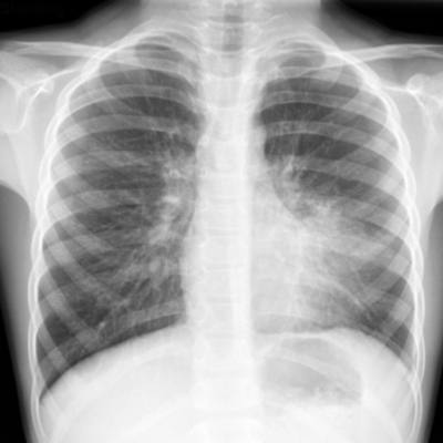
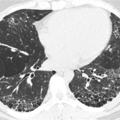
  2. Assessing Organ Involvement: Scleroderma doesn’t stop at the skin; it often targets vital organs such as the lungs, heart, and gastrointestinal tract. Imaging studies like chest X-rays, CT scans, and echocardiograms can provide vital information about the extent of organ involvement, helping doctors tailor treatment plans to individual patients.
  3. Disease Progression Monitoring: Scleroderma is a progressive disease, and regular monitoring is essential to track changes in organ function and tissue involvement. Imaging allows for ongoing assessment without invasive procedures, providing valuable data for treatment adjustments and long-term management.
  4. Evaluating Treatment Efficacy: Many treatment options are available to manage scleroderma, but their effectiveness varies from person to person. Imaging can help healthcare providers gauge how well treatment is working by tracking changes in organ function, tissue thickness, and inflammation levels.

Imaging plays a pivotal role in scleroderma research. By visualizing the disease at the cellular and molecular levels, scientists can gain deeper insights into its underlying mechanisms and develop more targeted therapies.

IAG’s team plays an active role in the scientific community and have led the development of novel scoring systems for early assessment of synovial inflammatory changes, quantification of inflammation (bone marrow oedema and synovial) and quantitative assessment of erosive changes.

About IAG, Image Analysis Group

IAG, Image Analysis Group is a strategic partner to bio-pharmaceutical companies developing new treatments to improve patients’ lives. Our dynamic Strategy, Trial Solutions and Bio-Partnering divisions work closely to meet critical needs of biotechnology companies: funding, clinical development, and monetization of their assets. We fuse decades of therapeutic insights, risk-sharing business model and agile culture to accelerate novel drug development. IAG broadly leverages its core imaging expertise, proprietary technology platform DYNAMIKA and capabilities to support an objective early go no/ go decision and drive excellence for tomorrow’s innovative therapeutic agents with speed.

Contact our expert team: imaging.experts @ ia-grp.com

READ NEXT CASE STUDY >
Experience: Scoring Systems
  • Eligibility and Safety Assessments
  • DAVIX (Digital Arteries Volume Index)
  • Quantitative Inflammation (DEMRIQ)
  • Organ Involvement (Xray or CT)
  • US-Doppler Quantification
Experience: Imaging
  • MRI
  • DCE-MRI
  • CT
  • X-ray
  • Ultrasound
Publications

Since 2007, over 2000 articles were published to cover scientific discoveries, technology break-throughs and special cases. We list here some critically important papers and abstracts.

Testimonials

Combining our technologies and business advisory services with promising life science companies has yielded spectacular results over the past five years. As a trusted partner to many biotech and pharma companies, IAG’s team is proud to share your words and quotes.

OSTEOPOROSIS

OSTEOPOROSIS

OSTEOPOROSIS

It is estimated that more than 200 million people are suffering from osteoporosis and according to the International Osteoporosis Foundation 1 in 3 women over the age of 50 years and 1 in 5 men will experience osteoporotic fractures in their lifetime.

The prevalence of osteoporosis among adults aged 50 and over increased from 9.4% in 2008 to 12.6% in 2018

A silent health condition is often only revealed in case of fracture. It’s a chronic, progressive disease of multifactorial etiology. It has been most frequently recognized in elderly white women, although it does occur in both sexes, all races, and all age groups.The implications on mortality and morbidity associated with osteoporotic fractures are significant; the mortality rate in an elderly person with a hip fracture approaches 20%.

Osteoporosis has a substantial economic burden on countries; the direct annual cost of treating osteoporotic fractures people is, on average, between 5.0 to 6.5 billion USD in Canada, Europe, and the USA alone, not taking into account indirect costs.

Prevention and treatment of osteoporosis is a massive challenge for Healthcare and Pharmaceutical industry.

… DXA

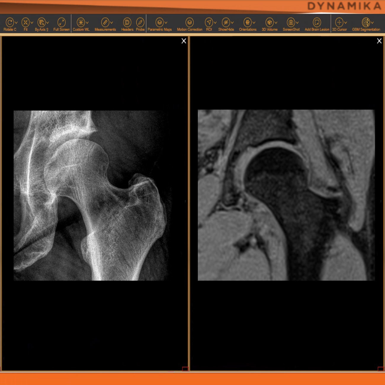
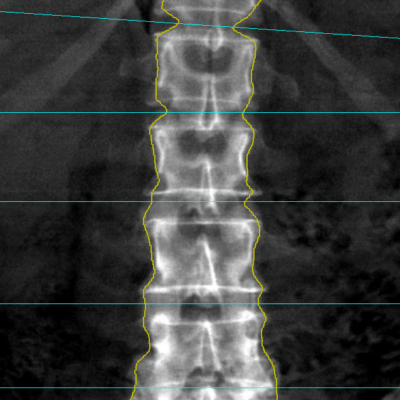
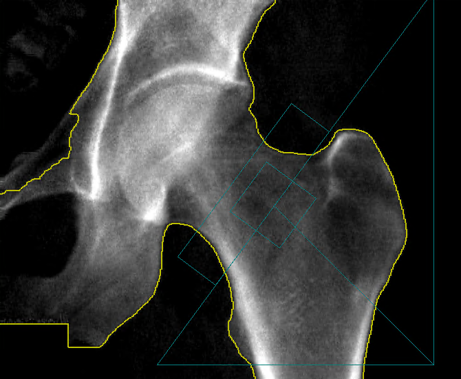
Osteoporosis [Porous Bone] is a systemic skeletal disease characterized by low bone density (Quantity) and microarchitectural deterioration of bone tissue (Quality), with a consequent increase in bone fragility. To measure the Bone Mineral Density (BMD) – DXA, also called dual-energy X-ray absorptiometry (DEXA), or bone densitometry, the “Gold-standard” for BMD measurement, the most accurate and standardized way to measure Bone Mineral Density (BMD).

As a mainly numerical imaging modality, DXA relies on the precision of its measurements, especially in clinical trials.

Dynamika allows seamless integration with the main DXA manufacturers enabling a central reading and a consistent outline for the Regions of Interest, essential for the accuracy of the results across different sites.
Able to use different reference populations for T-score calculations, Dynamika is a platform ready for DXA studies independently of its world location.

What is osteoporosis?

Osteoporosis or Low Bone Mass in Older Adults.

Economic burden of osteoporosis.

What is osteoporosis?

About IAG, Image Analysis Group

IAG, Image Analysis Group is a strategic partner to biopharmaceutical companies developing new treatments to improve patients’ lives. Our dynamic Strategy, Trial Solutions, and Bio-Partnering divisions work closely to meet the critical needs of biotechnology companies: funding, clinical development, and monetization of their assets. We fuse decades of therapeutic insights, a risk-sharing business model, and agile culture to accelerate novel drug development. IAG broadly leverages its core imaging expertise, proprietary technology platform DYNAMIKA and capabilities to support an objective early go no/ go decision and drive excellence for tomorrow’s innovative therapeutic agents with speed.

Contact our expert team: imaging.experts @ ia-grp.com

READ NEXT CASE STUDY >
Publications

Since 2007, over 2000 articles were published to cover scientific discoveries, technology break-throughs and special cases. We list here some critically important papers and abstracts.

Testimonials

Combining our technologies and business advisory services with promising life science companies has yielded spectacular results over the past five years. As a trusted partner to many biotech and pharma companies, IAG’s team is proud to share your words and quotes.

LUMBAR DISC DISEASE / SPINE

LUMBAR DISC DISEASE / SPINE

LUMBAR DISC DISEASE / SPINE

Lumbar disk disease may occur when a disc in the low back area of the spine bulges or herniates from between the bony area of the spine. Lumbar disk disease causes lower back pain and leg pain and weakness that is made worse by movement and activity. Degenerative disc disease in the lumbar spine, or lower back, refers to a syndrome in which age-related wear and tear on a spinal disc causes low back pain.

Low back pain (LBP) is the leading cause of disability in developed countries, with the number of people affected worldwide increasing annually.

Back pain is believed to originate from a multitude of lumbar spinal structures including ligaments, facet joints, vertebral periosteum, paravertebral musculature and fascia, blood vessels, annulus fibrosus, and spinal nerve roots.

Therefore, imaging of the lumbar spine has an established role in the evaluation of the LBP patient, and within the last few decades, almost every medical imaging modality has been applied in the evaluation of LBP.

Radiography

Radiography can visualize bony structures and can be used in suspected cases of traumatic, osteoporotic or pathologic vertebral fractures, malalignment, congenital defects, and late stages of inflammatory and infectious diseases. It is an inexpensive and we often recommend it in the initial screening of the spine.

In many of our studies, discography is applied to increase diagnostic accuracy for detecting painful disc levels in LBP with low false-positive rates.

Computed tomography (CT)

Computed tomography (CT) uses a 3D tomographic radiographic technique and is the standard of reference for imaging the vertebral bones, facet joints degeneration, and soft tissue calcification. Tissue contrast on CT is optimal in very dense structures such as bone. Dense soft tissues like tendons, ligaments, and annulus fibrosus can also, to some degree, be differentiated. This makes CT ideal for trauma imaging and fracture characterization in the spine.

We work with a number of CT and DUAL-energy CT (DECT) sites to ensure a new treatment impact has been captured through assessment of the morphology or the chemical composition of the different tissues. Some of the studies include multispectral CT scanners so that our clients have an opportunity to assess change sin bone marrow edema, ligaments, and tendons.

MRI of the lumbar spine

The usual MRI protocols of the lumbar spine include sagittal T1-weighted (T1w) turbo/fast spin echo (TSE/FSE), sagittal and axial T2-weighted (T2w), TSE/FSE images. Adding a sequence in the coronal plane at a later stage has been recommended to allow a better visualization of transitional vertebras, extraforminal disc herniations, extraforaminal nerve roots compression, degenerative vertebral translation, and paravertebral osteophytes.

The presence of fatty tissue in the bone marrow may obscure underlying pathologies on the T2w images without fat saturation. To overcome this issue, spectral fat suppression T2w or fluid sensitive such as the Short Tau Inversion Recovery (STIR) sequences have been used. In STIR sequences, fluid is bright because the contrast is a combination of T1w and T2w images. STIR is the most robust fat-suppression technique in the lumbar spine sensitive for detection of edema and may serve as a screening sequence for many neoplastic, infectious, and traumatic pathologies. However, it is less useful for degenerative conditions because of a high degree of noise and low resolution.

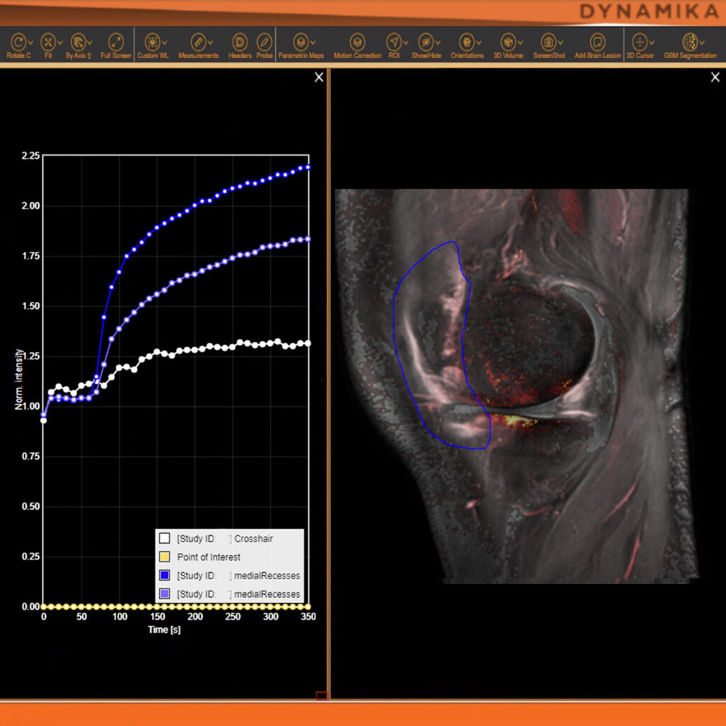
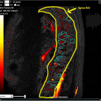
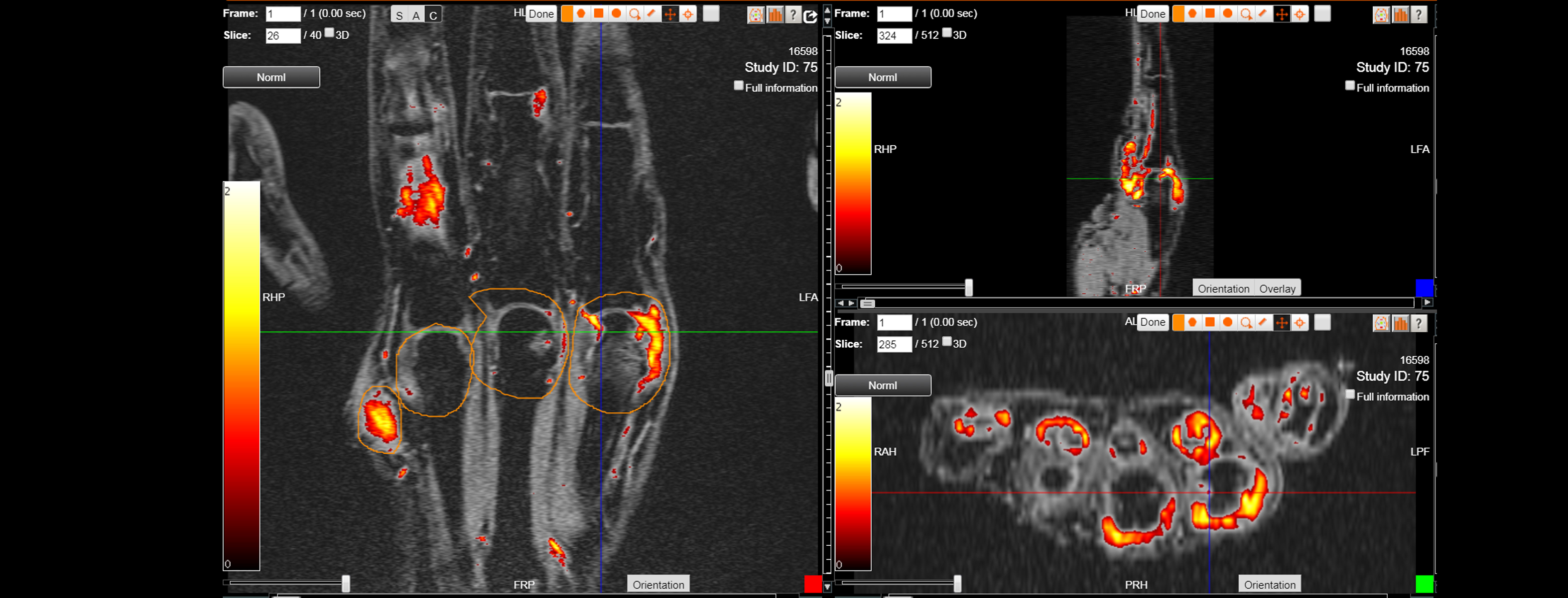
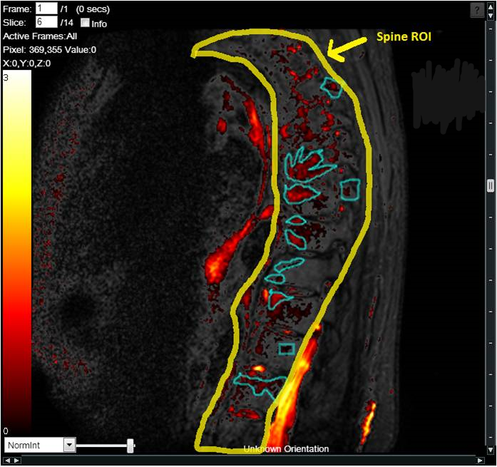
We also design protocols and assess changes using DIXON and DCE-MRI sequences, thus giving more precise information on the pain and inflammation.

IAG’s team has extensive experience in supporting Lower Back Pain, Disc Degeneration ad other Spine disease. Our team plays an active role in the scientific community and have led the development of novel scoring systems and formed strong collaborations with the industry’s Key Opinion Leaders as well as regulatory consultants and agencies.

We bring deep knowledge to help designing a trials, global experienced operations team and fully integrated cloud platform to support a trial that may require use of:

  • Xray
  • MRI, Dynamic Contrast Enhanced MRI, DIXON, STIR
  • CT, PET / CT

About IAG, Image Analysis Group

IAG, Image Analysis Group is a strategic partner to bio-pharmaceutical companies developing new treatments to improve patients’ lives. Our dynamic Strategy, Trial Solutions and Bio-Partnering divisions work closely to meet critical needs of biotechnology companies: funding, clinical development, and monetization of their assets. We fuse decades of therapeutic insights, risk-sharing business model and agile culture to accelerate novel drug development. IAG broadly leverages its core imaging expertise, proprietary technology platform DYNAMIKA and capabilities to support an objective early go no/ go decision and drive excellence for tomorrow’s innovative therapeutic agents with speed.

Contact our expert team: imaging.experts @ ia-grp.com


READ NEXT CASE STUDY >
Experience: Scoring Systems
  • MODIC
  • Disc morphology
  • Inflammation
  • Modified Pfirrmann score
  • Disc Height
  • Spinal Deformity
  • DEMRIQ
Experience: Imaging
  • X-ray
  • MRI
  • DCE-MRI
  • Ultrasound
  • CT
  • PET / CT
Publications

Since 2007, over 2000 articles were published to cover scientific discoveries, technology break-throughs and special cases. We list here some critically important papers and abstracts.

Testimonials

Combining our technologies and business advisory services with promising life science companies has yielded spectacular results over the past five years. As a trusted partner to many biotech and pharma companies, IAG’s team is proud to share your words and quotes.

GOUT

GOUT

GOUT

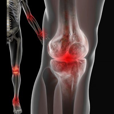
Gout is a form of arthritis — an inflammation of the joints — that causes sudden, severe attacks of pain, tenderness, redness, warmth, and swelling in some joints. Imaging modalities that have clinical relevance in gout include conventional radiography (CR), ultrasonography (US), computed tomography (CT), dual energy computed tomography (DECT), magnetic resonance imaging (MRI) and nuclear medicine.

In clinical trials, imaging biomarkers are used to assess joint inflammation and to be more specific about including a patient intro the trial, as imaging is more specific to and even pathognomonic of gout.

IAG’s team is actively working with drug developers to determine and help including into the trial inclusion / exclusion criteria, the right methodologies for patient stratification that can help differentiating gout from other inflammatory arthritis conditions. This especially critical for novel therapeutics targeting early disease.

IAG’s team has extensive expertise in using in CR, US and DECT to assist in monitoring response to treatment.

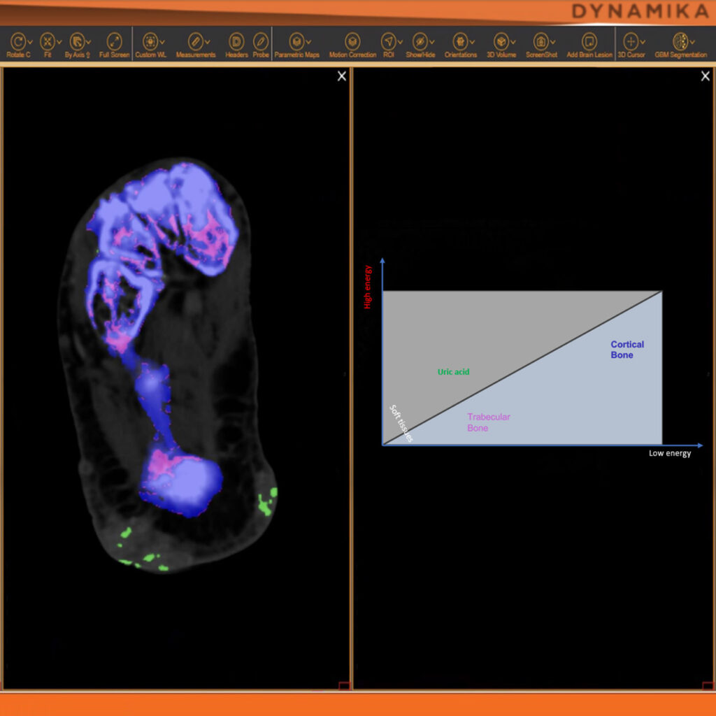
DECT provides good diagnostic accuracy for detection of monosodium urate (MSU) deposits in patients with gout. However, sensitivity is lower in patients with recent-onset disease. DECT can have a significant impact on clinical decision making when gout is suspected, but fails to demonstrate inflammatory potential of the MSU deposits.

IAG team is very active in scientific community and recent work was focused on completing an MRI trial assessing the response of sub-clinical, synovial inflammation to nine months of urate lowering therapy (ULT). Sub-clinical synovial-based inflammation has been documented in inter-critical gout. Together with academic and medical colleagues, we have investigated and reported a significant reduction in serum urate levels and when the MRI data was analysed with a quantitative method, we were able to score inter-critical synovitis in the MRI data in gout patients.

IAG team brings a great site network and significant experience in working with:

  • DECT
  • CT
  • MRI, inc Contrast MRI
  • US
  • PET

To support novel drug development for patients with gout. Reach out to our expert team, as you are designing and planning your trial.

About IAG, Image Analysis Group

IAG, Image Analysis Group is a strategic partner to bio-pharmaceutical companies developing new treatments to improve patients’ lives. Our dynamic Strategy, Trial Solutions and Bio-Partnering divisions work closely to meet critical needs of biotechnology companies: funding, clinical development, and monetization of their assets. We fuse decades of therapeutic insights, risk-sharing business model and agile culture to accelerate novel drug development. IAG broadly leverages its core imaging expertise, proprietary technology platform DYNAMIKA and capabilities to support an objective early go no/ go decision and drive excellence for tomorrow’s innovative therapeutic agents with speed.

Contact our expert team: imaging.experts @ ia-grp.com

READ NEXT CASE STUDY >
Experience: Scoring Systems
  • Gouty Arthritis MRI Score (GAMRIS)
  • DECT Assessment
  • US Assessment
  • Quantitative Inflammation (DEMRIQ)
Experience: Imaging
  • MRI
  • DCE MRI
  • CT
  • DECT
  • US
  • Conventional radiography
Publications

Since 2007, over 2000 articles were published to cover scientific discoveries, technology break-throughs and special cases. We list here some critically important papers and abstracts.

Testimonials

Combining our technologies and business advisory services with promising life science companies has yielded spectacular results over the past five years. As a trusted partner to many biotech and pharma companies, IAG’s team is proud to share your words and quotes.

LUPUS

LUPUS

LUPUS

Systemic lupus erythematosus (SLE) is an autoimmune disease that can affect multiple different organs, including the kidneys and central nervous system. With lupus, the body’s immune system targets its own body tissues. Lupus arthritis affects hands of the patients and lupus nephritis – the kidneys.
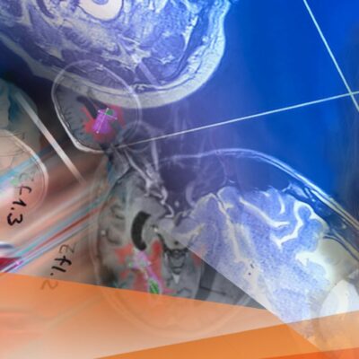
IAG’s team is supporting a number of academic and proof of concept trials that develop, test and validate various radiologic methods aiming to improve disease characterization in patients with SLE beyond simple anatomical endpoints.

In clinical trials, imaging biomarkers are used to assess joint inflammation and to be more specific about including a patient intro the trial, as imaging is more specific when added to the clinical assessment.

IAG’s team is actively working with drug developers to determine and help including into the trial inclusion / exclusion criteria, the right methodologies for patient stratification that can help differentiating lupus patients. This especially critical for novel therapeutics targeting early disease.

IAG’s team supported trials that involved various radiological examinations of patients with lupus, such as

  • Computed tomography (CT),
  • Magnetic Resonance Imaging (MRI),
  • Ultrasound (US).

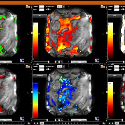
IAG’s team is active in scientific community and have contributed and actively led the developments in physiological non-contrast MRI protocols to assess tissue oxygenation, glomerular filtration, renal perfusion, interstitial diffusion, and inflammation-driven fibrosis in lupus nephritis (LN) patients. Pour radiology team worked on the vessel size imaging (VSI, an MRI approach utilizing T2-relaxing iron oxide nanoparticles) development and nuclear medicine team on the molecular imaging probes used to monitor the disease.

Reach out to our expert team, as you are designing and planning your trial.

About IAG, Image Analysis Group

IAG, Image Analysis Group is a strategic partner to bio-pharmaceutical companies developing new treatments to improve patients’ lives. Our dynamic Strategy, Trial Solutions and Bio-Partnering divisions work closely to meet critical needs of biotechnology companies: funding, clinical development, and monetization of their assets. We fuse decades of therapeutic insights, risk-sharing business model and agile culture to accelerate novel drug development. IAG broadly leverages its core imaging expertise, proprietary technology platform DYNAMIKA and capabilities to support an objective early go no/ go decision and drive excellence for tomorrow’s innovative therapeutic agents with speed.

Contact our expert team: imaging.experts @ ia-grp.com

READ NEXT CASE STUDY >
Experience: Scoring Systems
  • Visual MRI Scoring
  • Ultrasound Structural Erosion (ScUSSe)
  • Quantitative Inflammation (DEMRIQ)
Experience: Imaging
  • MRI
  • CT
  • US
  • PET
  • SPECT
Publications

Since 2007, over 2000 articles were published to cover scientific discoveries, technology break-throughs and special cases. We list here some critically important papers and abstracts.

Testimonials

Combining our technologies and business advisory services with promising life science companies has yielded spectacular results over the past five years. As a trusted partner to many biotech and pharma companies, IAG’s team is proud to share your words and quotes.

NEURO-MUSCULAR DISORDERS

NEURO-MUSCULAR DISORDERS

NEURO-MUSCULAR DISORDERS

Neuromuscular disorders affect the nerves that control voluntary muscles and the nerves that communicate sensory information back to the brain. Nerve cells send and receive electrical messages to and from the body to help control voluntary muscles. When the neurons become unhealthy or die, communication between the nervous system and muscles breaks down. As a result, muscles weaken and waste away (atrophy).

IAG’s experience with neuromuscular disorders include clinical development in:

  • Amyotrophic lateral sclerosis (ALS)
  • Multiple sclerosis
  • Muscular dystrophy
  • Myasthenia gravis
  • Myopathy
  • Myositis
  • Peripheral neuropathy
  • Spinal muscular atrophy

With the use of imaging biomarkers, our team helps to support a PI / physician in patient selection and disease diagnosis. As alternative to invasive methods, we recommend and help integrating into the trials Magnetic resonance imaging (MRI) scan of the brain, spinal cord, thigh or upper arm or Ultrasound (US), when appropriate.

MRI is a non-invasive imaging method, without ionizing radiation, which has the ability to visualize muscle, fat, connective tissue and bone. MRI has several advantages over muscle ultrasound, including that MRI has minimal operator-dependence and allows for excellent visualization of all muscles.

Added advantage of using MRI that we can provide the measurements for both volume and perfusion of muscle can be extracted from the scans.

Some of the studies that we support are investigating the disease changes at molecular level. IAG’s team has extensive experience in using PET / SPECT to reveal the metabolism of glucose within the bone and joint system.

IAG team is closely working with the recruiting sites and ensures seamless imaging data flow and assessment, to best support the development of novel treatments for these challenging conditions.

Our team is active in scientific community and our recent work investigates the association between pain and muscle perfusion, giving rise to many scientific collaborations. Our academic partners are working on the methodologies which allow for automated measurements of Fat Fraction and Muscle Volume. In any of our trials, the medical image analysis will be conducted with DYNAMIKA, that brings together quantitative methodologies for understanding treatment induced changes.

About IAG, Image Analysis Group

IAG, Image Analysis Group is a strategic partner to bio-pharmaceutical companies developing new treatments to improve patients’ lives. Our dynamic Strategy, Trial Solutions and Bio-Partnering divisions work closely to meet critical needs of biotechnology companies: funding, clinical development, and monetization of their assets. We fuse decades of therapeutic insights, risk-sharing business model and agile culture to accelerate novel drug development. IAG broadly leverages its core imaging expertise, proprietary technology platform DYNAMIKA and capabilities to support an objective early go no/ go decision and drive excellence for tomorrow’s innovative therapeutic agents with speed.

Contact our expert team: imaging.experts @ ia-grp.com

READ NEXT CASE STUDY >
Experience: Scoring Systems
  • Quantitative Assessment of Fat, Muscle
  • Lesion Volume
  • Texture Analysis
  • Total MR grading score
  • Fibro Fatty Infiltration
Experience: Imaging
  • MRI (T1, T2, DIXON)
  • Contrast MRI / DCE-MRI
  • STIR (Fat suppression sequences)
  • PET / SPECT
  • US
  • CT
Publications

Since 2007, over 2000 articles were published to cover scientific discoveries, technology break-throughs and special cases. We list here some critically important papers and abstracts.

Testimonials

Combining our technologies and business advisory services with promising life science companies has yielded spectacular results over the past five years. As a trusted partner to many biotech and pharma companies, IAG’s team is proud to share your words and quotes.

PSORIATIC ARTHRITIS

PSORIATIC ARTHRITIS

PSORIATIC ARTHRITIS

Psoriatic arthritis (PsA) is a heterogeneous inflammatory rheumatologic disorder characterized by peripheral arthritis, affecting 6% to 39% of patients suffering from psoriasis. PsA is classified as a seronegative spondyloarthropathy (SpA) because it shares certain features with other conditions included in that group. Indeed, spinal involvement has been reported in approximately 50% of patients with PsA. In addition, PsA is associated with enthesitis and dactylitis, which are extra-articular features common to SpA. Finally, the majority of patients with PsA are negative for the rheumatoid factor.

At the onset, the disease is typically oligoarticular, but can develop into asymmetrical polyarthritis. Merely 10% of patients present with articular and cutaneous changes simultaneously. In a half of patients, arthritis precedes psoriasis. In the initial stages of the disease, synovitis is usually observed in the knee, ankle, and metatarsophalangeal (MTP) joints and occurs in conjunction with dactylitis. Sternoclavicular joints are typically involved.

IAG’s team has extensive expertise and track record in supporting the development of novel therapeutics in PsA and SpA and helping to take these drugs from early phase all the way to phase 3 and approval. Currently IAG is supporting one of the largest and scientifically driven trials (MOSAIC). Our expertise is in use of

  • X-ray
  • MRI
  • US
  • CT

to support disease diagnosis and assessment of treatment response.

IAG’s team plays an active role in the scientific community and have led the development of novel scoring systems for early assessment of synovial inflammatory changes, quantification of inflammation and quantitative assessment of erosive changes.

Our extensive experience in using radiography, which is a conventional gold standard method for assessment of structural damage in spine and sacroiliac joints. We recommend and assure optimal use of Magnetic resonance Imaging (MRI) that has a number of advantages over plain radiography and ultrasound in assessing disease activity and damage in peripheral joints in PsA. MRI features of PsA include synovitis, dactylitis, tenosynovitis, erosions, bone edema and enthesitis. MRI and anatomic studies have helped researchers develop the concept of synovio-entheseal complex in the pathogenesis of PsA. IAG’s scientific team is actively developing and integrating into the trials novel sequences, including

  • Ultrashort T2 echo for assessment of entheses will help in understanding the similarities and differences between mechanical and inflammatory entheseal changes.
  • Whole-body MRI (WB MRI), with OMERACT as part of novel clinical development.
  • Dynamic contract enhanced MRI for quantitative assessment of inflammation.
  • Quantitative assessment of erosions.

About IAG, Image Analysis Group

IAG, Image Analysis Group is a strategic partner to bio-pharmaceutical companies developing new treatments to improve patients’ lives. Our dynamic Strategy, Trial Solutions and Bio-Partnering divisions work closely to meet critical needs of biotechnology companies: funding, clinical development, and monetization of their assets. We fuse decades of therapeutic insights, risk-sharing business model and agile culture to accelerate novel drug development. IAG broadly leverages its core imaging expertise, proprietary technology platform DYNAMIKA and capabilities to support an objective early go no/ go decision and drive excellence for tomorrow’s innovative therapeutic agents with speed.

Contact our expert team: imaging.experts @ ia-grp.com

READ NEXT CASE STUDY >
Experience: Scoring Systems
  • Psoriatic Arthritis MRI Scoring (PsAMRIS)
  • DEMRIQ, DCE-MRI
  • Exploratory: WB-MRI
Experience: Imaging
  • X-ray
  • Ultrasound
  • DCE-MRI
  • MRI
Publications

Since 2007, over 2000 articles were published to cover scientific discoveries, technology break-throughs and special cases. We list here some critically important papers and abstracts.

Testimonials

Combining our technologies and business advisory services with promising life science companies has yielded spectacular results over the past five years. As a trusted partner to many biotech and pharma companies, IAG’s team is proud to share your words and quotes.

ANKYLOSING SPONDYLITIS

ANKYLOSING SPONDYLITIS

ANKYLOSING SPONDYLITIS

Ankylosing Spondylitis (AS), a form of spondylarthritis (SpA), is a systemic inflammatory rheumatic disease. Although the exact pathogenesis of AS is not yet fully understood, as with rheumatoid arthritis, the swelling, pain, and joint damage that characterize AS are considered to be the result of complex autoimmune and inflammatory processes based on both environmental and genetic factors.

IAG’s team has extensive expertise and track record in supporting the development of novel therapeutics in AS and SpA and helping to take these drugs from early phase all the way to phase 3 and approval. Our expertise is in use of

  • X-ray
  • MRI
  • Dynamic Contrast Enhanced MRI
  • CT

to support disease diagnosis and assessment of treatment response.

IAG’s team plays an active role in the scientific community and have led the development of novel scoring systems for early assessment of synovial inflammatory changes, quantification of inflammation and quantitative assessment of erosive changes. Our extensive experience in using radiography, which is a conventional gold standard method for assessment of structural damage in spine and sacroiliac joints. Our medical science team have worked with drug developers in trials incorporating Computed Tomography (CT), which is a more sensitive method for assessment of structural changes in the spine and sacroiliac joints.

In many trials, early diagnosis of sacroiliitis, the most frequent clinical symptom often accompanied by inflammatory back pain, and other inflammatory lesions of the spine such as spondylitis and spondylodiscitis, can be visualized early by magnetic resonance imaging (MRI), especially using fat saturating STIR or Gd-Enhanced Dynamic MRI sequences. IAG’s team helped to integrate ultrasonography and MRI into the trials for visualization and quantification of peripheral arthritis and enthesitis.


About IAG, Image Analysis Group

IAG, Image Analysis Group is a strategic partner to bio-pharmaceutical companies developing new treatments to improve patients’ lives. Our dynamic Strategy, Trial Solutions and Bio-Partnering divisions work closely to meet critical needs of biotechnology companies: funding, clinical development, and monetization of their assets. We fuse decades of therapeutic insights, risk-sharing business model and agile culture to accelerate novel drug development. IAG broadly leverages its core imaging expertise, proprietary technology platform DYNAMIKA and capabilities to support an objective early go no/ go decision and drive excellence for tomorrow’s innovative therapeutic agents with speed.

Contact our expert team: imaging.experts @ ia-grp.com

READ NEXT CASE STUDY >
Experience: Scoring Systems
  • New York criteria
  • Stoke Ankylosing Spondylitis Spinal Score (SASSS)
  • Bath Ankylosing Spondylitis Radiology Index (BASRI)
  • MRI Spine Score
Experience: Imaging
  • MRI
  • DCE-MRI
  • X-ray
  • CT
  • US
Publications

Since 2007, over 2000 articles were published to cover scientific discoveries, technology break-throughs and special cases. We list here some critically important papers and abstracts.

Testimonials

Combining our technologies and business advisory services with promising life science companies has yielded spectacular results over the past five years. As a trusted partner to many biotech and pharma companies, IAG’s team is proud to share your words and quotes.